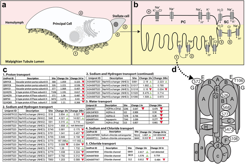
Fig 2. Changes in phosphorylation of ion channels, pumps, and aquaporins.
a. Illustration of the two major cell types in the Malpighian tubule epithelium: principal cells (PCs) are larger and have an apical brush border of microvilli filled with mitochondria containing V-type H+ ATPase pumps and other ion transporters; stellate cells (SCs) are smaller and participate in Cl- and water transport. Numbers correspond to different regions where particular transport mechanisms occur, and correspond to the numbers in parts b and c. b. Ion channels and aquaporins associated with transcellular transport are illustrated. c. Specific amino acid residues on proteins with phosphorylation changes of at least 1.5-fold between unfed and blood-fed mosquitoes are shown in white ovals. The numbers in the figure correspond to tables where proteins with related functions are grouped. Tables contain the Uniprot IDs for each protein as well as the fold-change in phosphorylation of each residue between unfed and either 1hr PBM or 24hr PBM mosquitoes. Green arrows represent an increase of at least 1.5-fold in phosphorylation at the specified residue between unfed and PBM mosquitoes, and red arrows represent a decrease of at least 1.5-fold in phosphorylation at the specified residue between unfed and PBM mosquitoes. Numbers of each table correspond to transport mechanisms illustrated in parts a and b. d. V-type H+ ATPase model adapted from Zhao et al (2015) and Beyenbach & Piermarini (2011) showing the location of subunits with differential phosphorylation. Subunits are shown in gray, and specific phosphorylated residues are denoted with white ovals.

