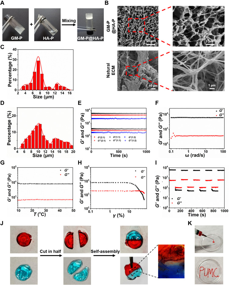
Fig. 3. Structure, mechanical, and self-healing properties of the GM-P@HA-P hydrogel.
(A) Representative optical pictures for preparing the GM-P@HA-P hydrogel. (B) Representative SEM images of lyophilized GM-P@HA-P hydrogel and natural cutaneous ECM prepared by spray drying. (C and D) Diameter distribution diagram of pores in the GM-P@HA-P hydrogel (C) and natural skin ECM (D). Data represent means ± SDs (n = 3). (E to H) Rheological analysis of GM-P@HA-P hydrogel as a function of time (E), angular frequency (F), temperature (G), and shear strain (H). (I) The self-healing property of GM-P@HA-P hydrogel. (J) Macroscopic self-healing test of GM-P@HA-P hydrogel. Dyes for hydrogel staining were rhodamine and aniline blue for red and blue color, respectively. (K) Optical images of GM-P@HA-P hydrogel demonstrating the injectability.

