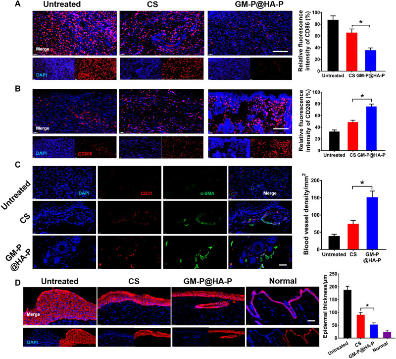
Fig. 8. GM-P@HA-P hydrogel promoted M2-type macrophage polarization and angiogenesis during the wound healing.
(A and B) Representative immunofluorescence images and quantification analysis of macrophage phenotype in the regenerated skin tissues on day 21 after diabetic wound healing. Macrophages were stained with CD86 (A) or CD206 (B) in red, and the nuclei were stained with 4′,6-diamidino-2-phenylindole (DAPI) in blue. Scale bar, 100 μm. (C) Representative immunofluorescence images of CD31 (red)/α-SMA (green) staining in the regenerated skin tissues and quantification of blood vessel density on day 21 after wound healing. Nuclei were stained with DAPI in blue. Scale bar, 50 μm. (D) Representative immunofluorescence images for CK 14 staining to show the epidermal thickness. The epithelial cells were stained with CK 14 in red, and the nuclei were stained with DAPI in blue. Scale bar, 50 μm. *P < 0.05.

