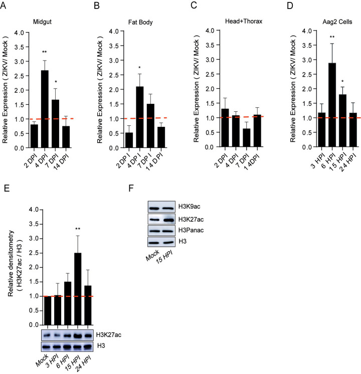
Fig 2. ZIKV infection modulates the expression and activity of AaCBP.
Aag2 cells as well as mosquitoes were infected with ZIKV at a MOI of 2.0, and 60 PFUs, respectively. Mosquito infections were performed by intrathoracic injections. The expression of AaCBP in mosquitoes (A-C) or Aag2 cells (D) was measured by qRT-PCR on the indicated tissues and days post infection. E and F. Western blot of total protein extract from Aag2 cells infected with ZIKV virus, or mock-infected at different time points. Monoclonal antibodies against H3K9ac, H3K27ac, H3 panacetylated (Panac), or H3 (as a loading control) were used. The intensity of the bands was quantified by densitometry analysis plotted as a graph using ImageJ (NIH Software). Western blotting was performed on 3 independent biological replicates and one representative is shown here. Error bars indicate the standard error of the mean; statistical analyses were performed by Student’s t test. *, p <0.05; **, p <0.01.

