
Figure 1.
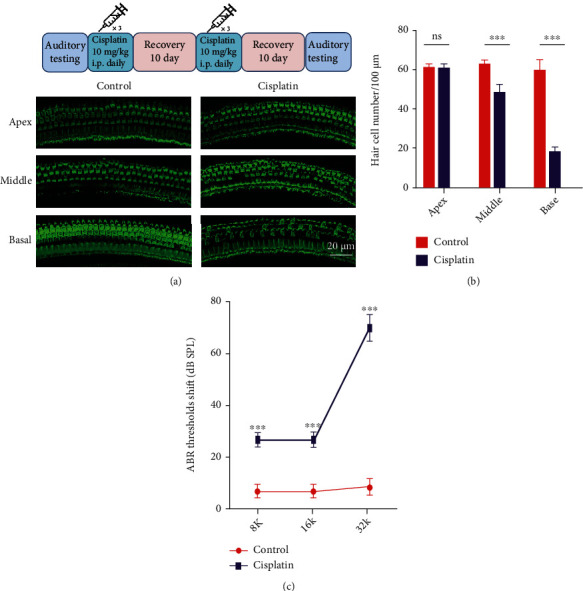
Cisplatin-induced cell death in the mouse cochlea. (a, top) Mode of cisplatin administration to CBA/J mice. (a, bottom) Phalloidin-labeled hair cells of the organ of Corti. (b) Hair cell quantification and statistical analysis. (c) Statistical analysis of the ABR threshold shift. ∗p < 0.05, ∗∗p < 0.01, and ∗∗∗p < 0.001. Scale bar, 20 μm.