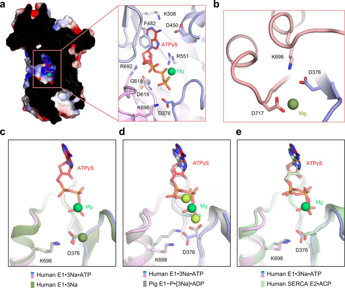
Fig. 3. ATP binding modes in NKA.
a ATP binding pocket between the N and P domains and the key residues around ATPγS (red). b Mg2+ binding site and the phosphorylation site (Asp376) in the E1·3Na state. c Comparison of the Mg2+ binding site and the phosphorylation site (Asp376) between the E1·3Na (dark green) and E1·3Na·ATP (blue to magenta) states. d, e Comparison of the ATP binding pocket between the E1·3Na·ATP (blue to magenta) and E1~P·[3Na]·ADP (grey) or SERCA E2·ACP (pale green) states. Mg2+ are coloured forest green, green and pea green in E1·3Na, E1·3Na·ATP and E1~P·[3Na]·ADP, respectively.

