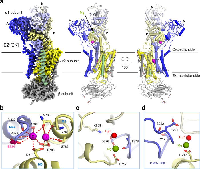
Fig. 5. Overall structure of E2·2[K].
a The overall cryo-EM map of E2·2[K] is shown in the left panel, and two perpendicular views of the overall structure are shown in the middle and right panels. The α1 subunit is rainbow coloured from blue for N-terminus to yellow for the C-terminus; the β1 subunit is coloured grey; the γ2 subunit is coloured pale green. b In the E2·2[K] state, two K+ are bound to transmembrane K+ binding sites (I and II). c Mg2+ stabilizes phosphorylation site (Asp376). d The hallmark 219TGES motif in the A domain is located near Mg2+. K+ are coloured magenta; Mg2+ is coloured olive green; and water molecules are coloured red. The glycosylation moieties are shown as sticks. e extracellular, c cytosolic, M transmembrane helix.

