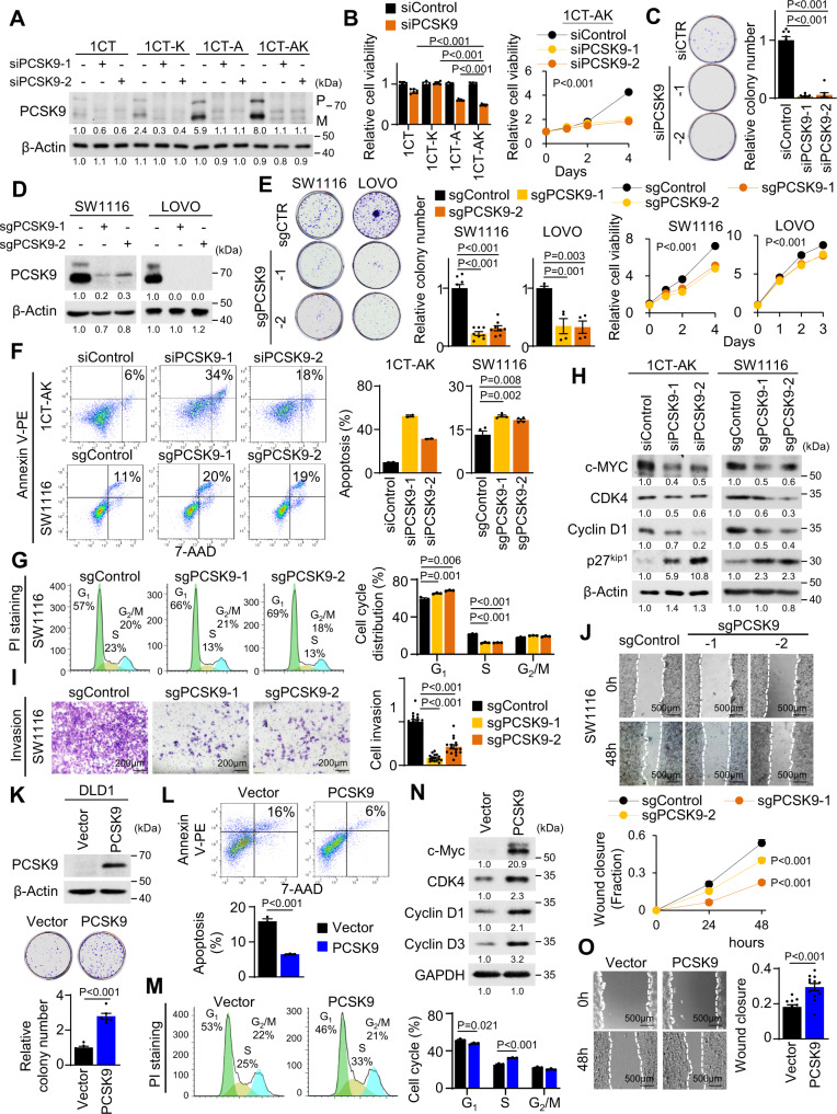
Fig. 2. PCSK9 promotes malignant phenotypes in APC/KRAS-mutant CRC.
A PCSK9 knockdown in 1CT isogenic cell lines. B PCSK9 knockdown suppressed cell proliferation (n = 6) and C colony formation (n = 8) in 1CT-AK cells. D PCSK9 knockout in SW1116 and LOVO cells. E PCSK9 knockout reduced cell growth (n = 6) and colony formation (SW1116: n = 8, 7 days; LOVO: n = 4, 14 days) in SW1116 and LOVO cells. F PCSK9 knockdown (72 h) or knockout induced cell apoptosis (1CT-AK: n = 2; SW1116: n = 4) and G inhibited G1-S cell cycle progression (n = 3). H Western blot revealed that PCSK9 knockdown/knockout induced the expression of cleaved PARP and p27Kip1, while suppressing c-Myc, CDK4 and Cyclin D1. I PCSK9 knockout suppressed Matrigel invasion (72 h, 16 replicates in 4 independent experiments) and J wound healing closure (12 replicates in 4 independent experiments) in SW1116 cells. K PCSK9 overexpression in DLD1 cells increased cell proliferation (n = 6, 7 days), L inhibited apoptosis (n = 3) and M promoted G1-S progression (n = 3). N PCSK9 increased CDK4, Cyclin D1, and Cyclin D3, but suppressed p27Kip1 in DLD1 cells. O PCSK9 overexpression promoted cell migration in DLD1 cells (48 h, 12 replicates in 4 independent experiments). Data shown are means of biological replicates; ± SEM (B, C, E–G, I–M, O). Two-tailed Student’s t-test (B, C, E–G, I, K–M, O). Two-tailed two-way ANOVA (growth curves in B, E, J). Source data are provided as a Source Data file.

