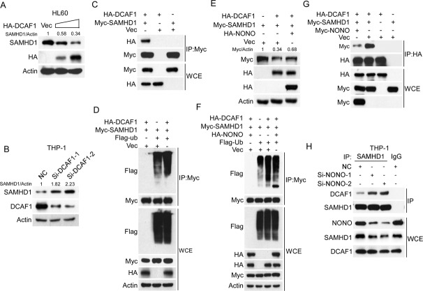
Fig. 4. NONO blocks the ubiquitination-mediated degradation of SAMHD1 by DCAF1.
A, B Overexpression or silencing of DCAF1 decreased or increased SAMHD1 expression, respectively. A HL60 cells were transfected with increasing concentrations of HA-tagged DCAF1 24 h, and the levels of the indicated proteins were determined by western blotting. B THP-1 cells were transfected with DCAF1 siRNAs for 24 h, and the levels of the indicated proteins were then determined by western blotting. C DCAF1 interacts with SAMHD1. 293 T cells were transfected with HA-tagged DCAF1 and Myc-tagged SAMHD1 for 24 h. Co-IP was performed with anti-Myc agarose, and the interactions were examined by western blotting. D Overexpression of DCAF1 increased the ubiquitination level of SAMHD1. Myc-tagged SAMHD1 was cotransfected with HA-tagged DCAF1, Flag-ubiquitin or empty vector into 293 T cells for 24 h, and the ubiquitination level of SAMHD1 was determined using IP followed by western blotting. E NONO blocked DCAF1-mediated degradation of SAMHD1. Myc-tagged SAMHD1 and HA-tagged DCAF1 were cotransfected into 293 T cells with or without overexpression of HA-tagged NONO for 24 h. The levels of the indicated proteins were determined by western blotting. F Overexpression of NONO inhibited DCAF1-mediated ubiquitination of SAMHD1. Myc-tagged SAMHD1, HA-tagged DCAF1, and Flag-tagged ubiquitin were cotransfected into 293 T cells with or without overexpression of HA-tagged NONO for 24 h. The ubiquitination of SAMHD1 was evaluated by IP followed by western blotting. G NONO inhibits the interaction of DCAF1 and SAMHD1. Myc-tagged SAMHD1 and HA-tagged DCAF1 were cotransfected into 293 T cells with or without overexpression of Myc-tagged NONO for 24 h. Cells were subsequently subjected to co-IP followed by western blotting with the indicated antibodies. H Silencing of NONO increases the interaction of DCAF1 with SAMHD1. THP-1 cells silencing NONO for 48 h were lysed and were subsequently subjected to co-IP followed by western blotting with the indicated antibodies. For respective immunoblots, the protein levels were quantified by ImageJ software.

