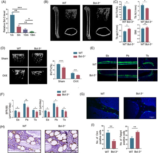
FIGURE 1.

Deletion of Bcl‐3 exacerbates bone loss and reduces bone formation. (A) Bcl‐3 mRNA in femurs from 1 m, 3 m, 10 m and 18 m wild‐type (WT) male mice was assessed by qRT‐PCR. (N = 3 independent experiments). (B) Representative micro‐computed tomography (μCT_ images of femurs from 10‐week‐old Bcl‐3–/– and WT male mice. (N = 3 mice/group). (C) Quantitative measurements of BV/TV, Tb.N, Tb.Sp and SMI in 10‐week‐old Bcl‐3–/– and WT mice by μCT. (N = 3 mice/group). (D) Representative μCT images of femurs from 10‐week‐old Bcl‐3–/– and WT female mice following sham and ovariectomy (OVX). (N = 3 mice/group). (E,F) Representative images of calcein double labelling of endosteal bone (Eb), periosteal bone (Pb) and trabecular bone (Tb) with quantification of BFR/BS and MAR in the femurs of 10‐week‐old WT and Bcl‐3–/– male mice. (G) Representative immunostaining of OCN in 10‐week‐old Bcl‐3–/– and WT male mouse femurs. Scale bar, 50 μm. (N = 3 mice/group). (H) Representative immunostaining of FABP4 in 6‐month‐old Bcl‐3–/– and WT male mouse femurs. Scale bar, 100 μm. (N = 3 mice/group). (I) Quantification of immunostaining of OCN and FABP4. The data are presented as the mean ± SD. *P <.05; **P <.01; ***P <.005; ****P <.0001 vs. control group. Statistical analysis was performed using Student's t‐test (A, C, F and I) and two‐way ANOVA test (D). Primary antibodies: OCN, Abcam(ab93876); FABP4, Abcam(ab92501)
