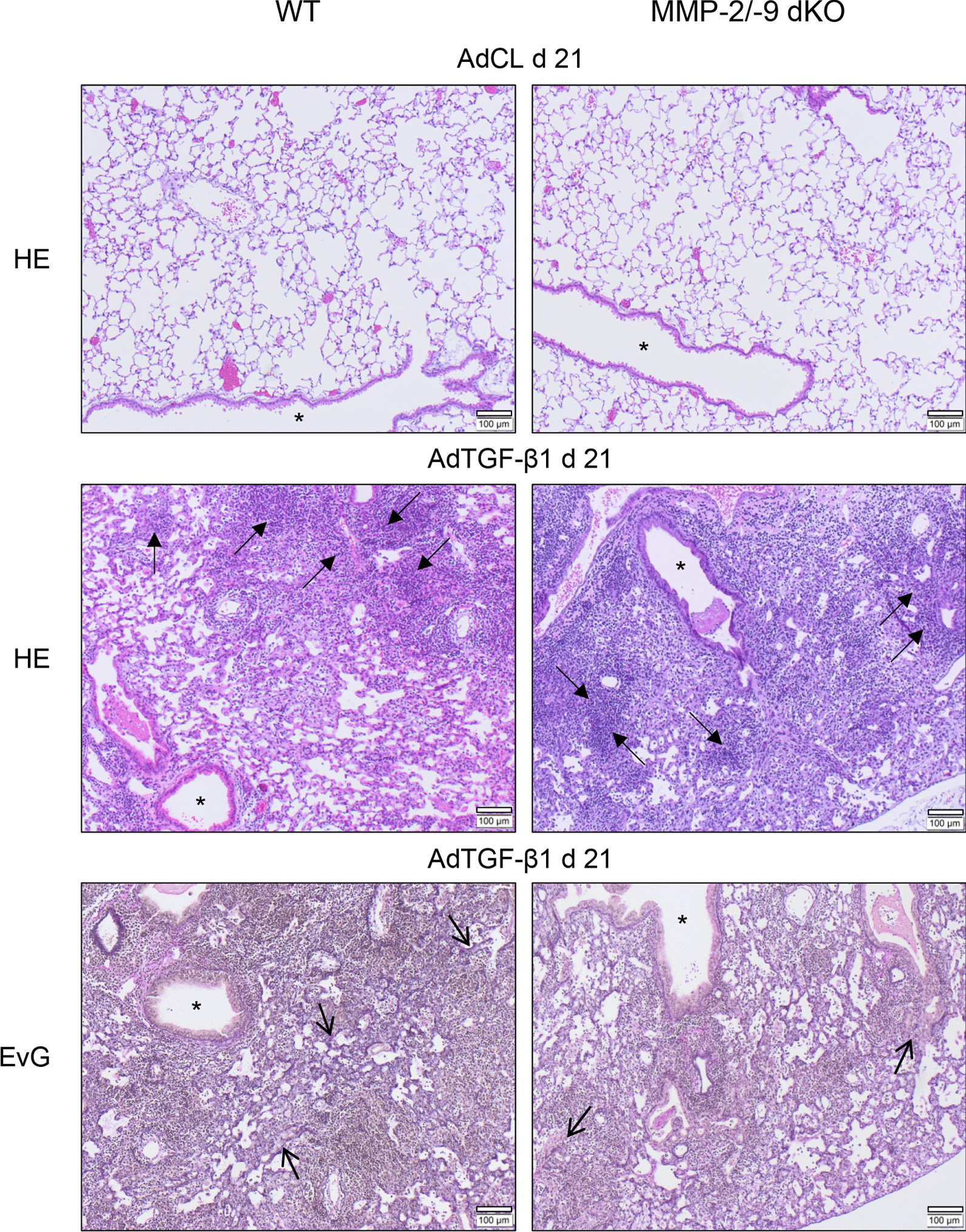
Fig. 4.

Lung histopathology of WT and MMP-2/-9 dKO mice with AdTGF-β1-induced lung fibrosis. WT and MMP-2/-9 dKO mice were exposed to AdCL vector or AdTGF-β1 for 21 days. Subsequently, lung sections were stained with hematoxylin/eosin or Elastica van Gieson (EvG) for assessment of lung tissue remodeling. The provided histology is representative of n = 4 mice per experimental group. Closed arrows, lymphoplasmacellular infiltrates; open arrows, areas of increased fibrotic remodeling; asterisk, bronchus (original magnification, × 10; scale bar, 100 µm)
