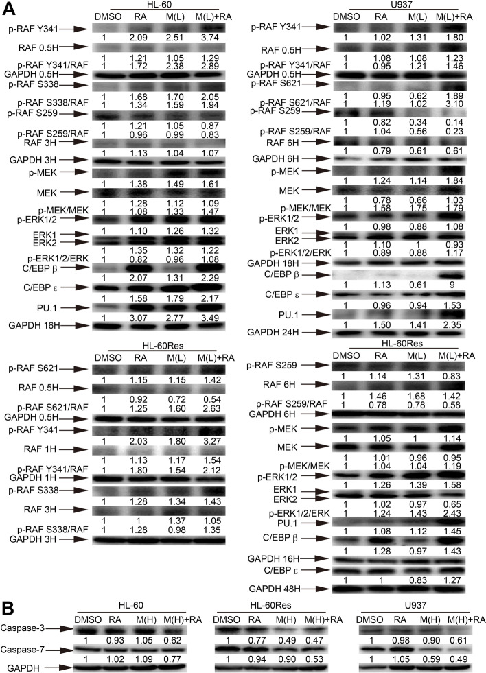
Fig. 2.
Mido(H)-ATRA activates caspase-3/7, and mido(L)-ATRA activates RAF-MEK-ERK and increases the expression of C/EBPs and PU.1. (A) HL-60 cells were treated with 0.25 μM midostaurin (M(L)) and/or 0.1 μM ATRA (RA). U937 and HL-60Res cells were treated with 0.1 μM midostaurin (M(L)) and/or 1 μM ATRA (RA). Changes in protein expression were detected at different time points and the corresponding expression of GAPDH at each time point was used as the internal control. (B) HL-60 cells were treated with 0.5 μM midostaurin (M(H)) and/or 0.1 μM ATRA (RA) for 3 h. U937 and HL-60Res cells were treated with 0.5 μM midostaurin (M(H)) and/or 1 μM ATRA (RA) for 12 and 24 h, respectively. GAPDH was used as the internal control. Most of the membranes were cut prior to hybridization and the original blots are presented in Supplementary Fig. 3. The values shown below each lane indicate relative units, with the values in DMSO-treated cells defined as 1.0. The phosphorylated protein/unphosphorylated protein ratios are also shown

