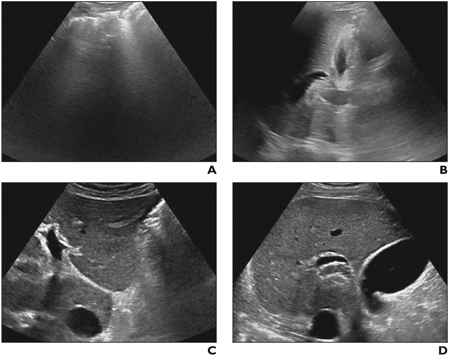
Fig. 2—

Difference in visualization score between scan locations in 52-year-old man with nonalcoholic steatohepatitis–related cirrhosis. Examinations were performed by high-volume sonographers using same scanner model and interpreted by community radiologists.
A and B, Transverse gray-scale ultrasound image of left lobe (A) at initial liver ultrasound in emergency department shows complete obscuration of liver by bowel gas. Longitudinal image of right lobe (B) shows parenchyma largely obscured by lung and rib. Visualization score C was assigned.
C and D, Gray-scale images of left (C) and right (D) lobes 2 months later when patient underwent liver ultrasound as outpatient show marked improvement in image quality. Visualization score A was assigned.
