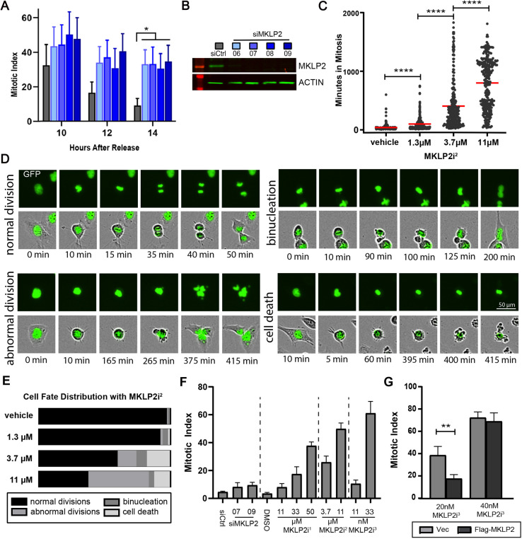
Fig. 1.
MKLP2 deficiency leads to prolonged mitosis. (A) Mitotic index (percentage of cells in mitosis) in HeLa cells transfected with the siRNAs indicated in B after release from DTB. *P<0.05, n>200 cells/condition from two independent experiments (two-way ANOVA with Tukey post-hoc test). (B) Western blot of representative MKLP2 knockdown from A. (C) Dose-dependent effect of MKLP2i2 on duration of mitosis in HeLa histone H2B–GFP cells ****P<0.0001, n>200 cells/condition from three independent experiments (unpaired two-tailed t-test). (D) Images portraying cell fates following treatment with MKLP2i2 for analysis quantified in C and E. Top rows, histone H2B–GFP channel; bottom rows, phase contrast and GFP merge. (E) Percentage of cells from C and D with indicated fates at the corresponding MKLP2i2 treatment dose. (F) Mitotic index in HeLa cells 14 h after release from DTB in the presence of various doses of MKLP2i1, MKLP2i2, MKLP2i3 and siMKLP2 (n=50 cells/experiment from three independent experiments). (G) Graph depicting mitotic index of HeLa cells transfected with either pCS2 flag (empty vector) or pCS2 MKLP2 in between DTB. Cell were treated with either 20 nM MKLP2i3 or 40 nM MKLP2i3 6 h before entering mitosis. The mitotic index was quantified 14 h after release from DTB. **P<0.01, n=50 cells/experiment from two independent experiments (unpaired two-tailed t-test). Results in A, F and G are mean±s.e.m. Mean is highlighted in C.

