Fig. 3.
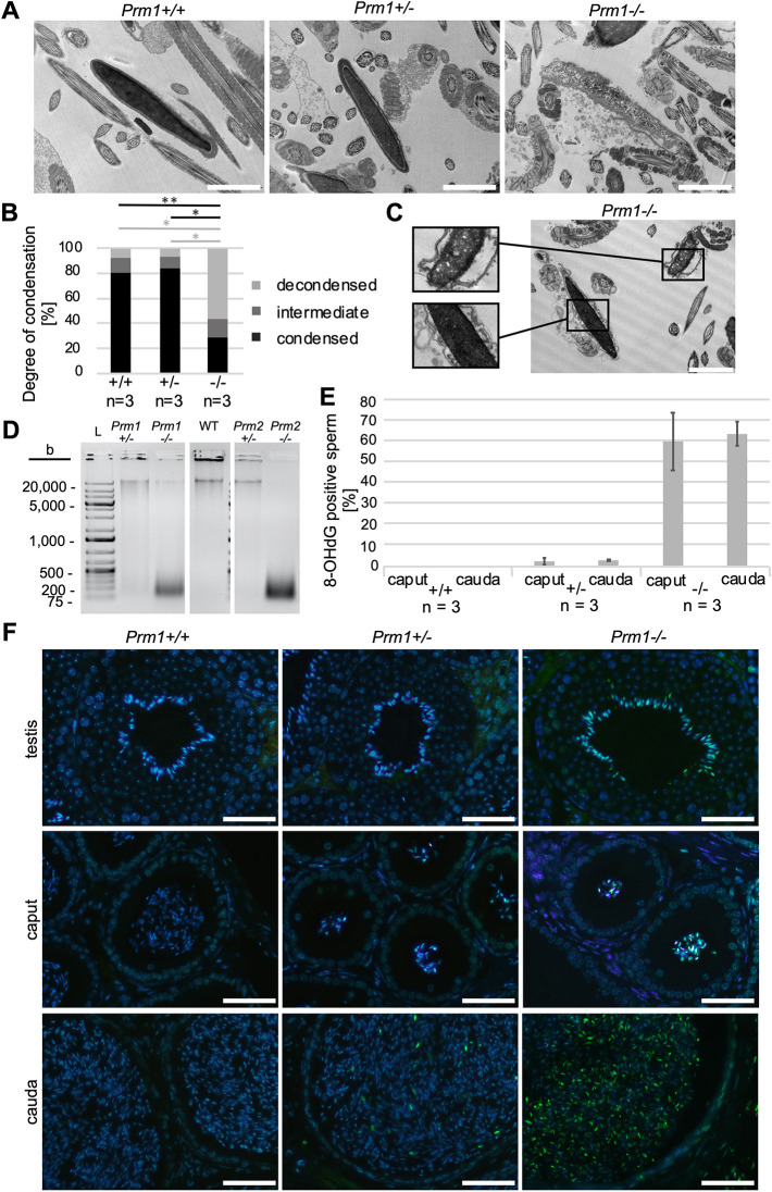
Analysis of chromatin condensation and ROS-induced DNA damage in epididymal Prm1-deficient sperm. (A) Representative transmission electron micrographs of Prm1+/+, Prm1+/− and Prm1−/− epididymal sperm. (B) Quantification of DNA condensation of epididymal sperm from Prm1+/+, Prm1+/− and Prm1−/− males (n=3); 100 sperm per male were analyzed. (C) Transmission electron micrograph of Prm1−/− epididymal sperm. (D) Agarose gel loaded with genomic DNA isolated from epididymal sperm of Prm1+/−, Prm1−/−, Prm2+/−, Prm2−/− and WT males separated by electrophoresis. Additional lanes loaded with ladders (L) were cut from the image. (E) Percentage of 8-OHdG-positive sperm on tissue sections of caput and cauda epididymis of Prm1+/+, Prm1+/− and Prm1−/− mice (n=3). (F) Representative IF staining against 8-OHdG in testis, caput epididymis and cauda epididymis tissue sections from Prm1+/+, Prm1+/− and Prm1−/− males. Data are mean±s.d. and were analyzed using a two-tailed, unpaired Student's t-test (*P<0.05; **P<0.005). Scale bars: 2 µm in A,C; 50 µm in F.

