Fig. 5.
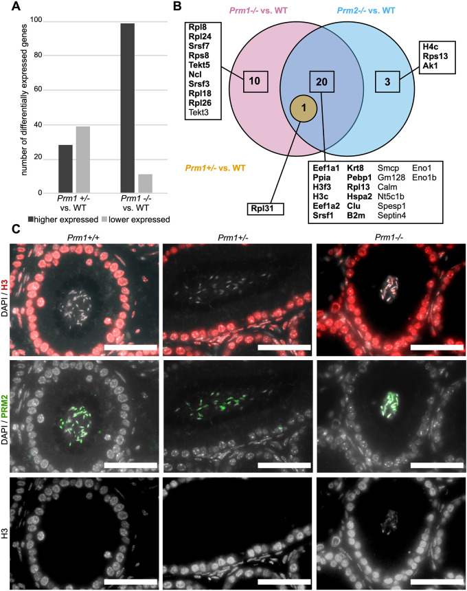
Differentially expressed genes in the testis and altered protein abundances in sperm in protamine-deficient males. (A) Number of differentially expressed genes subdivided into higher and lower expressed genes in testis of Prm1+/− and Prm1−/− males compared with WT males. (B) Venn diagram illustrating changes in abundances of proteins from sperm basic protein extractions of Prm1−/−, Prm1+/− and Prm2−/− males compared with WT. Proteins that were more abundant are in bold. Non-bold proteins showed lower abundance compared with WT. (C) IHC stainings against histone H3 (red) and PRM2 (green) of Prm1+/+, Prm1+/− and Prm1−/− caput epididymal tissue sections. DAPI (in gray) was used as the counterstain. The H3 stainings are additionally shown as single gray channel pictures. Scale bars: 50 µm.

