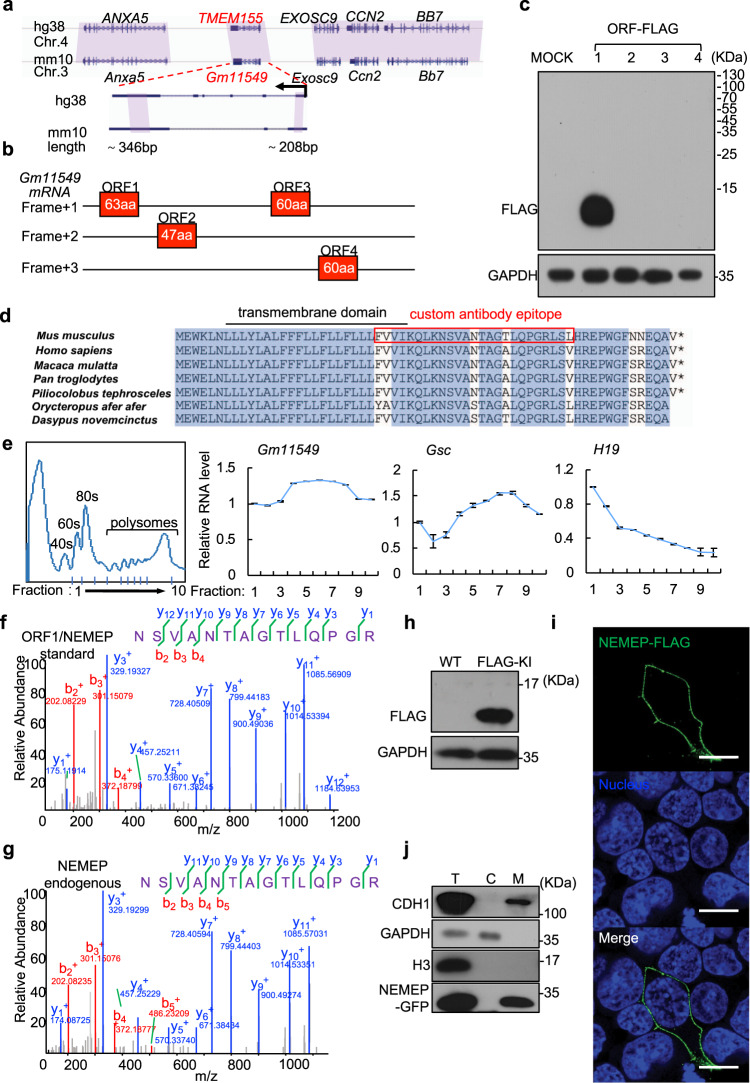
Fig. 2. Gm11549 encodes a transmembrane micropeptide: NEMEP.
a Conservation of the Gm11549 and TMEM155 loci between mouse (Mus musculus) and human (Homo sapiens). The purple shadow marks the conserved regions between mouse and human. The expanded region shows the conserved sequences between Gm11549 and TMEM155. b Predicted ORFs in the Gm11549 RNA sequence. c HEK293T cells were transiently transfected with expression vectors for all four of the predicted ORFs, each tagged with a FLAG epitope at their C termini. Immunoblotting analysis for accumulation of proteinaceous gene products from the predicted ORFs, detected with an anti-FLAG antibody. GAPDH was used as internal control. d Amino acid sequence alignment of ORF1 in the indicated species. e Ribosome profiling. Cytosolic lysates from differentiated cells were subjected to sucrose gradient centrifugation to isolate fractions including free 40/60 S subunits, monosomes, di/trisomes, and polysomes. RNAs were then extracted from these fractions and the Gm11549, Gsc, and H19 levels were quantified by qPCR. Gsc served as the controls for coding transcripts and H19 was the control for noncoding transcripts. f Following pull-down using FLAG antibody, a targeted proteomics analysis of lysates from transiently transfected HEK293T cells expressing C-terminal FLAG-tagged ORF1/NEMEP: MS/MS spectrum of one unique peptide corresponding to the Gm11549 ORF1 protein (henceforth: “NEMEP” for Nodal Enhanced MEsendoderm microPeptide). g MS/MS spectrum for one unique peptide corresponding to NEMEP from a targeted proteomics analysis following pull-down using a NEMEP polyclonal antibody raised against a 25-residue region of the NEMEP from adult mouse brain tissue lysates. h Immunoblotting analysis of NEMEP-3xFLAG in differentiated WT and NEMEP-3xFLAG knock-in mESCs with anti-FLAG antibody. GAPDH was used as internal control. i Immunofluorescence detection of NEMEP-FLAG in NEMEP-FLAG expressing HEK293T cells with anti-FLAG antibody (green). Nuclei are stained with Hoechst33342 (blue). Original magnifications, 10 µm. j Subcellular localization of NEMEP-GFP in NEMEP-GFP overexpression mESCs. Total cell lysates (T), Cytosol (C), and membrane fractions (M) from NEMEP-GFP overexpression mESCs were assessed by immunoblotting against specific markers and GFP. The GAPDH, Histone H3, and CDH1 protein respectively served as markers for the cytosolic, nuclear, and membrane fractions. c, e, f–j: All data are representative of three independent experiments with similar results. Source data are provided as a Source Data file.

