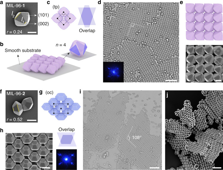
Fig. 5. Truncation-dependent assembly of MIL-96 2D films.
a–e Square (tp) lattice formed by MIL-96-1. SEM image (a) of MIL-96-1 with small truncation (r = l1/l2 = 0.24); the (101) and (002) facets are labeled. Cartoon in b shows each MIL-96-1 particle adheres to the substrate by (101) facet and packs with four neighboring particles by contacting their trapezoidal faces (purple) (n = 4). Cartoons in c show the unit cell and the antiparallel face overlap. Optical microscope image (d), laser diffraction pattern (d, inset), cartoon and SEM image (e) show the square lattices assembled from MIL-96-1. f–j Centered rectangular (oc) lattice formed by MIL-96-2. SEM image of MIL-96-2 (r = 0.52) is shown in f. Cartoons (g), SEM (h), and optical microscope image (i) show the structure of MIL-96-2 superlattice (axial angle = 108°). The unit cell is labeled in g. The face overlap and laser diffraction pattern are shown in (h). SEM image in j shows the pieces of MIL-96-2 films. Scale bars: 0.5 μm (a, e, f, h), 5 μm (d, i), and 2 μm (j).

