Fig. 1.
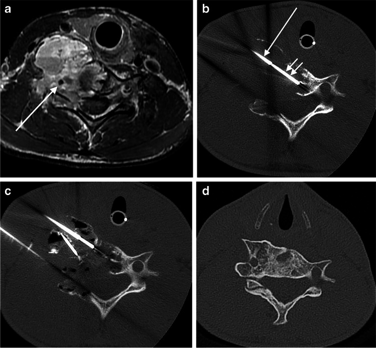
Cross-sectional images from a 14-year-old boy (patient 9) with a C6 aneurysmal bone cyst (ABC). a Axial pre-treatment supine T1-W MR image following contrast administration shows a destructive and exophytic multilocular cystic mass replacing the right half of the C6 ring and surrounding the vertebral artery (arrow). b Axial supine CT image during biopsy and first treatment at the same level as (a) shows a 14-gauge (G) guiding needle (single arrow) through which passes a 15-G biopsy needle (double arrows). c Axial supine CT on the same date and at the same level as in (a and b) shows three separate needles within different portions of the ABC with doxycycline foam (appearing black from air in foam) throughout the different loculations of the lesion. d Diagnostic axial CT at same level 3 years after last treatment shows healing of the ABC

