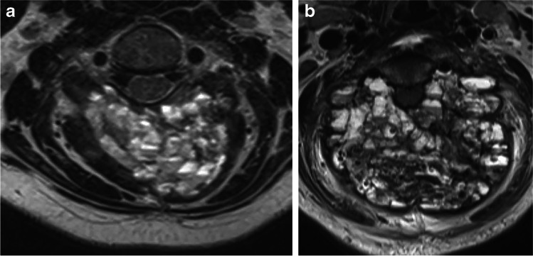
Fig. 3.
Axial MR images in a 10-year-old girl (patient 13) with an aneurysmal bone cyst (ABC) of the posterior half of the C3 ring. a Axial MR image 2 weeks before the first treatment shows expansion and replacement of spinous process and bilateral lamina at C3. More aggressive ABCs tend to have innumerable tiny cysts, as depicted in this case. b Axial MR image at the same level 3 months later shows near doubling in size of this very aggressive lesion, now with effacement of the canal

