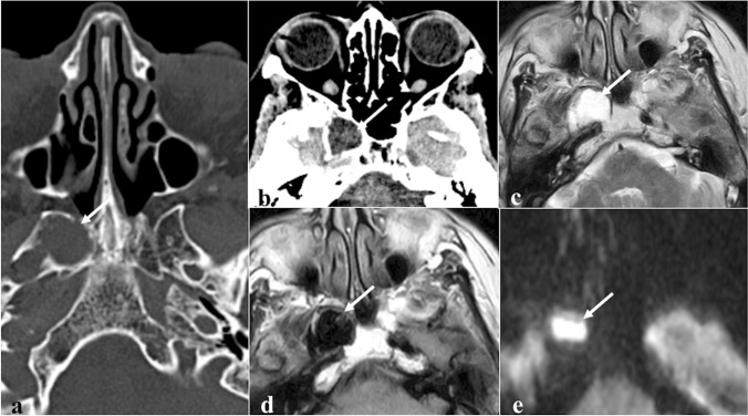
Fig. 4.
Sphenoid epidermoid cyst as an incidental finding in a 74-year-old female patient. Axial CT images (a and b) reveal a rounded lytic lesion in the right greater sphenoid wing (white arrows) with sclerotic margins and homogenous density similar to cerebrospinal fluid. Axial MRI images show that the lesion has high SI on T2W (c), heterogeneously low/dirty SI on fluid attenuated inversion recovery (d), and high SI on DWI b1000 sequences (e) due to the restricted water movements. Epidermoid cyst has similar features as arachnoid cyst on CT. Arachnoid cysts would have demonstrated homogeneous low SI on fluid attenuated inversion recovery MRI — as low as cerebrospinal fluid — and facilitated diffusion on DWI

