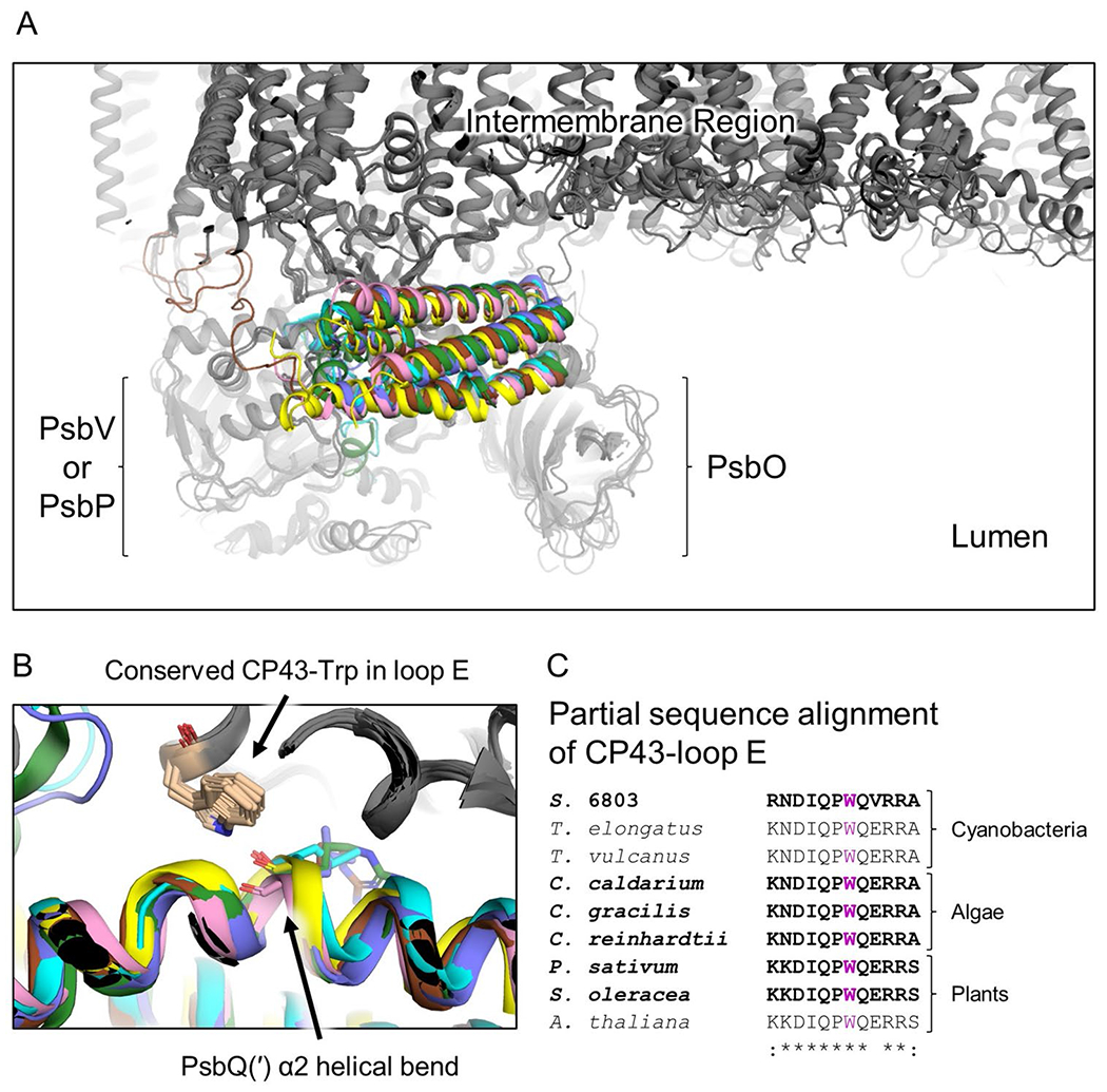
Fig. 3.

Binding site of PsbQ(′) and conserved CP43-Trp interaction. A Superimposed PsbQ(′)-containing PSII structures. Structures are colored grey except the PsbQ(′) subunit where that from Synechocystis 6803 is yellow, C. caldarium is pink, C. gracilis is brown, C. reinhardtii is green, P. sativum is blue, and S. oleracea is cyan. The regions of PsbO and PsbV/PsbP, the intermembrane region of the protein complexes, and the lumenal space, are labeled. B Focus on the PsbQ(′)-CP43 interface where a conserved CP43-Trp sidechain donates a H-bond to the backbone of α2 in its helical bend of each PsbQ(′) subunit. PsbQ(′) is colored as in A. C Partial sequence alignment of the CP43-loop E showing the conservation of the Trp residue (purple) in B. Bold sequences correspond to those structures shown in A and B, which are those PSII structures with associated PsbQ(′) subunits. The Clustal Omega (Sievers et al. 2011) sequence similarity identifiers are shown below the sequence alignment. Synechocystis 6803 is abbreviated S. 6803
