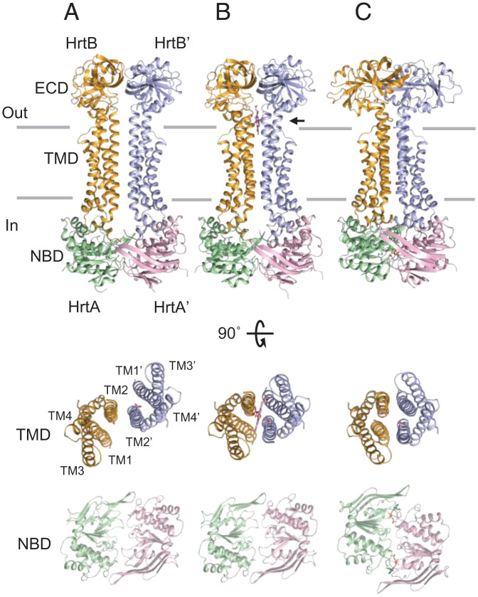
Fig. 2.
Crystal structures of HrtBA. (Top) (A) Unliganded form. (B) Heme-bound form. (C) AMPPNP-bound form. (Middle) Top views of the TM helices of the HrtB dimer. The aa (1 to 12, 249 to 263, 332 to 343) are omitted. (Bottom) Top views of the HrtA subunits. Glu219 of HrtB, heme, and AMPPNP are indicated by stick models. Horizontal lines indicate the hydrophobic layer of the membrane. An arrow indicates heme.

