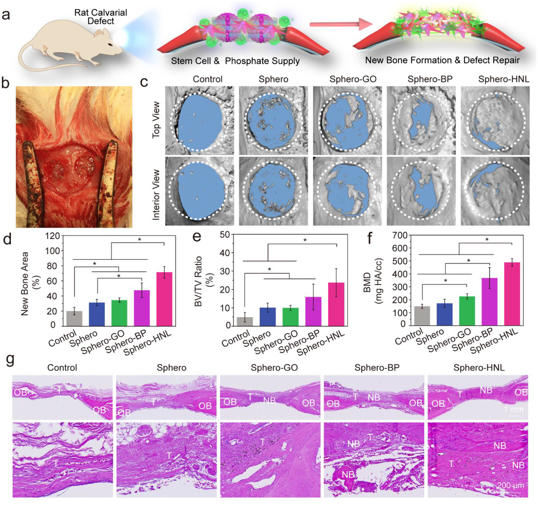
Fig. 5.

In vivo bone formation in rat calvarial defects. a) Schematic demonstration of bone formation in defects treated with stem cell spheroids and b) photograph of in vivo implantation of the spheroids. c) Micro-CT reconstruction of the rat cranial defect only (Control) or defects with stem cell spheroids after 8 weeks of implantation. Quantitative analysis of d) bone area, e) bone volume/tissue volume ratio, and f) bone mineral density. g) H & E staining of tissue samples from rat cranial defects without (Control) or with stem cell spheroids. NB: New Bone; OB: Original Bone; T: Tissue. (*: p < 0.05).
