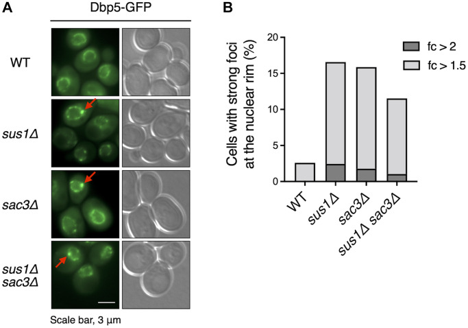
Figure 6.
Dbp5 is mislocalized in sus1Δ cells. (A) Fluorescence microscopy analysis of Dbp5-GFP in WT cells and the indicated mutants. The left and right panels show GFP and DIC images, respectively. Red arrows indicate strong foci at the nuclear rim. (B) The percentage of cells containing strong Dbp5-GFP foci shown in (A). The ratio of the GFP intensity of each strong focus to that in the outer region of the strong focus was calculated. Light and dark gray color bars in the graph indicate the percentage of cells with >1.5- and 2-fold of the focus ratio, respectively.

