
Figure 2 |. Light Microscopy Metadata is essential for the assessment, interpretation, reproducibility, comparison, and re-use of the results of microscopy experiments.
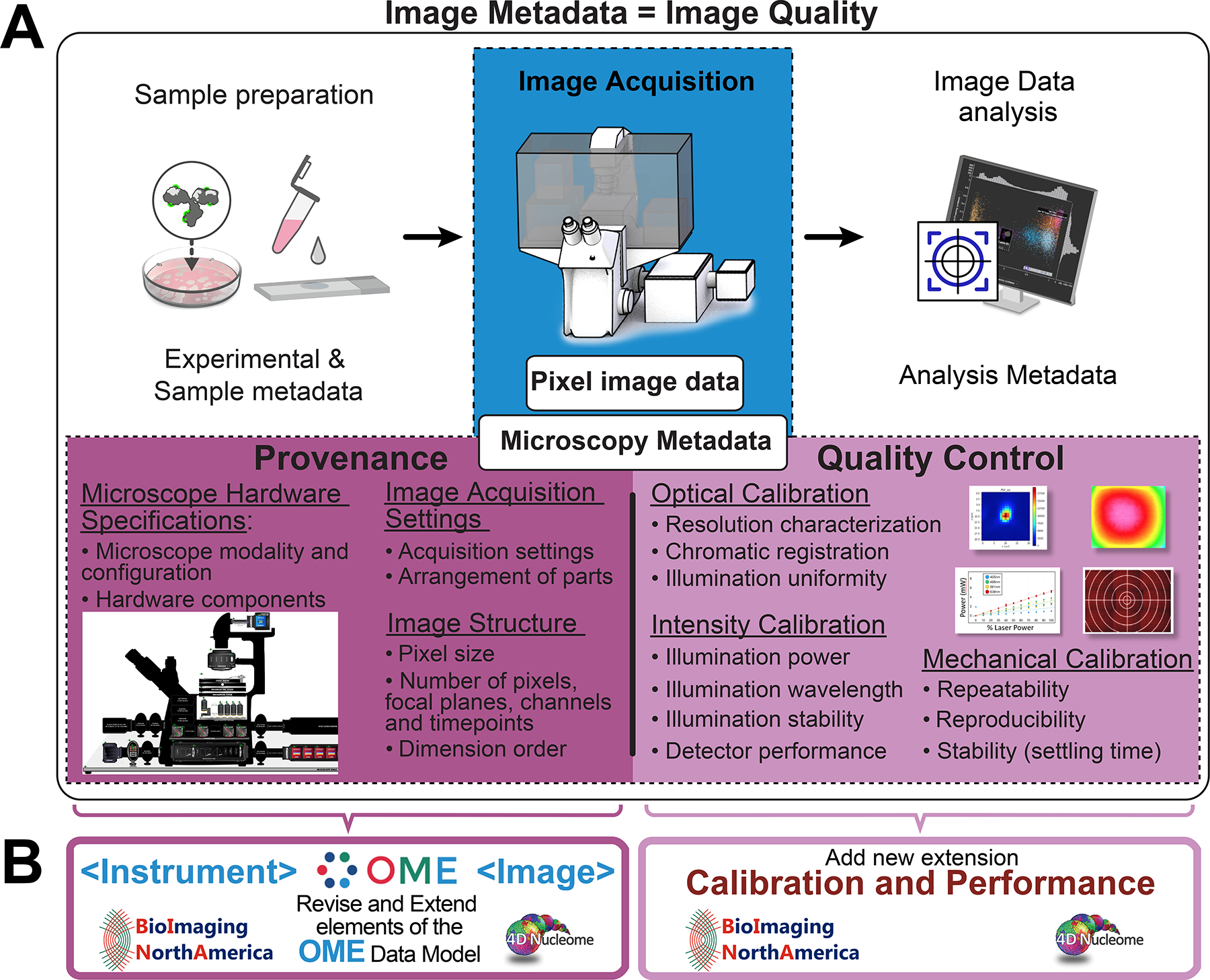
A) Depicted is a schematic representation of a typical bio-imaging experiment and of the Image Metadata that has to be collected to ensure the quality reproducibility and scientific value of the resulting Pixel Image Data (blue box). Specifically, imaging experiments and the associated metadata can be subdivided as follows: 1) Sample preparation documented by Experimental & Sample Metadata; 2) Image Data Acquisition documented by Microscopy Metadata; 3) Image Analysis documented by Analysis Metadata. In turn, Microscopy Metadata can be subdivided in two categories as indicated (magenta boxes): 1) Provenance metadata includes information that documents Microscope Hardware Specifications, Image Acquisition Settings, and Image Structure; 2) Quality Control metadata includes metrics that quantitatively assess the performance of the microscope and the quality of image data and are obtained through the execution of specifically designed Optical, Intensity and Mechanical calibration procedures. B) In order to capture and store Microscopy Metadata, the 4DN-BINA-OME specifications presented here take advantage of the structure of the OME Data Model4,5, which serves as the de facto specification for the exchange of image data and metadata. Specifically, Provenance metadata is stored into revised and extended versions of the <Instrument> and <Image> elements of the OME Data Model. On the other hand, Quality Control metadata is stored utilizing a newly designed Calibration and Performance extension of the same model.