FIG. 3.
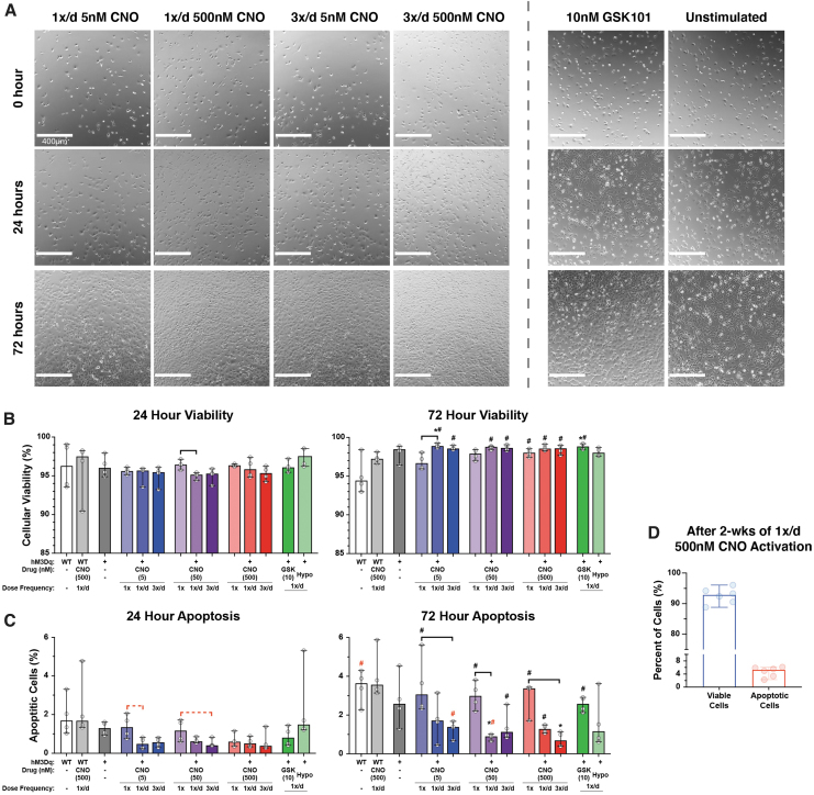
Effect of CNO stimulation on hM3Dq cell proliferation and health during early stimulatory culture. (A) Location-matched DIC images revealed CNO concentration and frequency-dependent increases in the proliferative capacity of hM3Dq cells; elevated stimulatory pressures induced increased cell proliferation compared with less-frequent and lower concentration stimulation; however, all CNO stimulated groups appeared to proliferate faster than the non-CNO groups. (B) Cell viability was quite consistent among all groups after 24 h of culture, at >95% cells. At 72 h, only unstimulated WT cells experienced a—minor—decrease in viability, dropping from 96% to 94%; no significant changes in viability were observed among WT or unstimulated hM3Dq cells; and hM3Dq cells multiply stimulated with CNO or GSK101 saw significant increases in viability at 72 h compared with 24 h, increasing by ∼2–3% percentage points. (C) Apoptosis for all groups increased at 72 h compared with 24 h, but this increase was significantly suppressed in multiply stimulated hM3Dq cells in a concentration- and frequency-dependent manner; the highest CNO-stimulated group (3 × /day 500 nM) displayed <1% apoptosis. (D) Viability and apoptosis in hM3Dq cells stimulated once daily with 500 nM CNO was very high (>92%) and low (<5%), respectively, after 2 weeks of culture. *p < 0.05 compared with WT (& WT+CNO), Kruskal–Wallis test with Dunn's multiple-corrections tests. Bar = p < 0.05 for the comparison between indicated groups for the noted CNO concentration, Kruskal–Wallis test with Dunn's multiple-correction tests. #p < 0.05 for comparison between respective 72- and 24-h outcomes, Mann–Whitney test. Statistical comparisons indicated in red are those that fell just short of significance, p = 0.0558 for Kruskal–Wallis test and p = 0.0571 for Mann–Whitney test. DIC, differential interference contrast.

