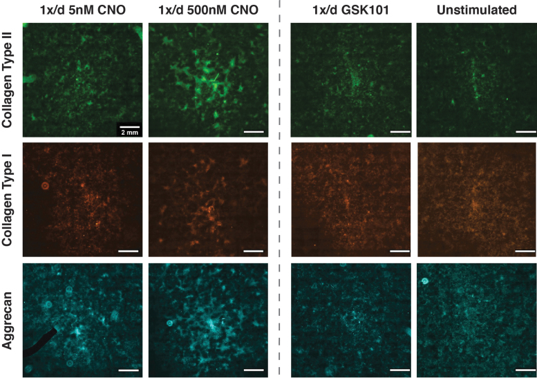
FIG. 6.
ICC staining revealed that CNO-mediated activation of hM3Dq enhanced collagen type II and aggrecan deposition. CNO stimulation resulted in the qualitative appearance of concentration-dependent enhancement of antibody staining for collagen type II (top row) and aggrecan (bottom row). Once-daily stimulation with 500 nM CNO exhibited the highest levels of deposition of both proteins, whereas 5 nM CNO displayed increased staining relative to GSK101 and unstimulated cultures; appreciable but reduced staining for collagen type I (middle row) was also detected. The black region in the 1 × /day 5 nM CNO aggrecan image is a “masked-out” fluorescent dust particle.

