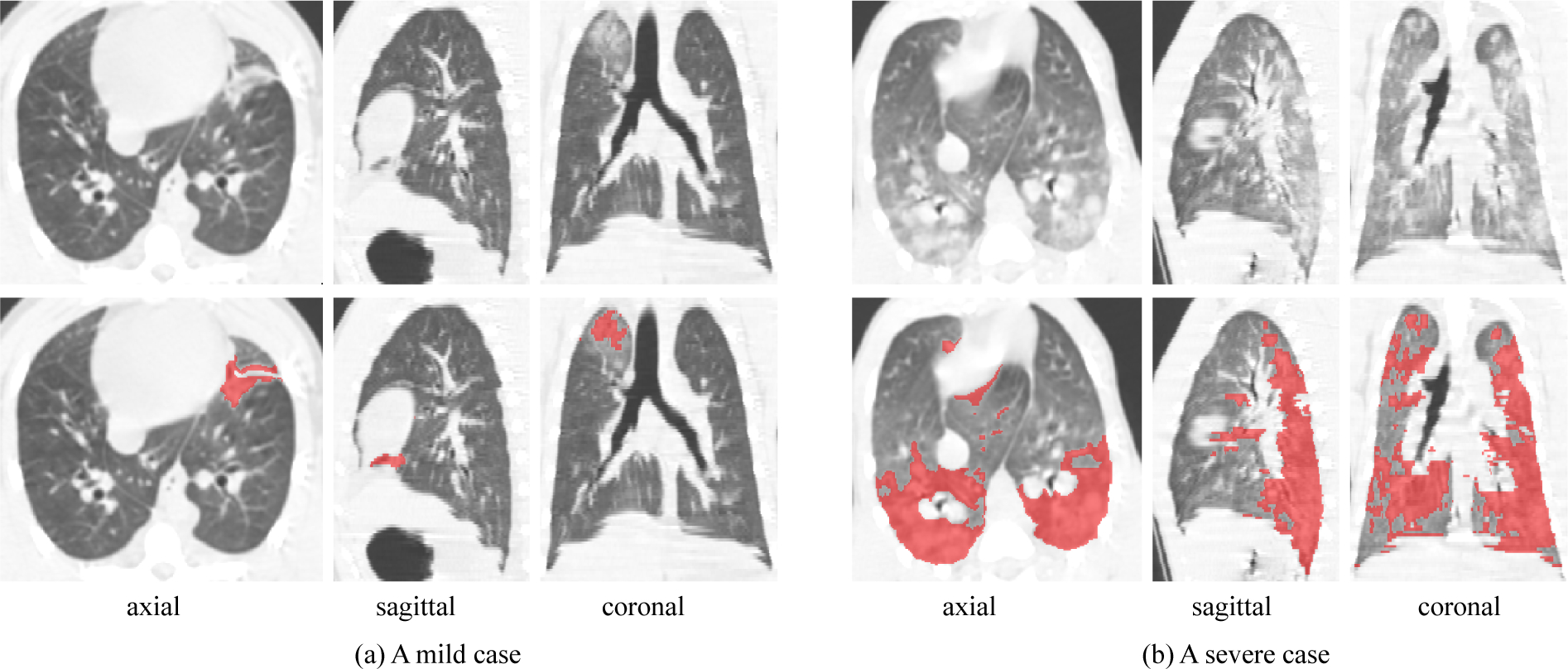
Fig. 1:

Examples of radiation-induced Pulmonary Fibrosis (PF) of the Rhesus Macaque. The first row shows lung CT images, and the second row shows manual segmentation results of PF lesions. Note that the ambiguous boundary, irregular shape and various size and position make the segmentation task challenging.
