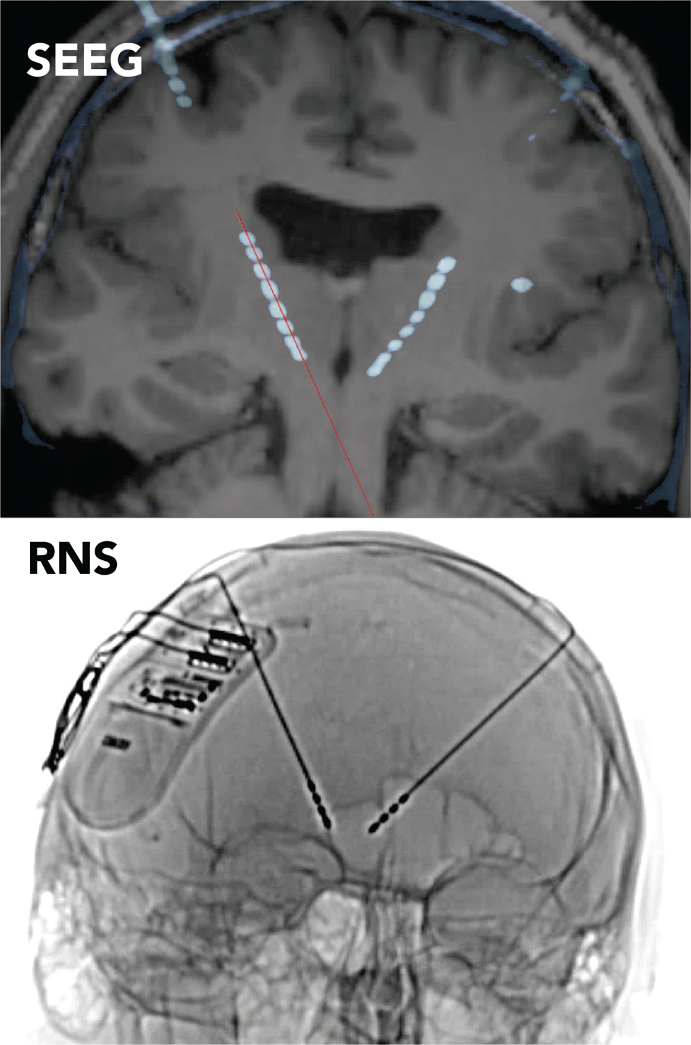
Figure 3. Thalamic implantation during SEEG.

Post-SEEG implantation CT fused to the pre-operative brain MRI, demonstrating the position of the fronto-thalamic SEEG leads (top). The orientation and SEEG lead trajectories chosen for this patient were the same as that planned for use with transfrontal implantation of bilateral thalamic leads for RNS (bottom).
