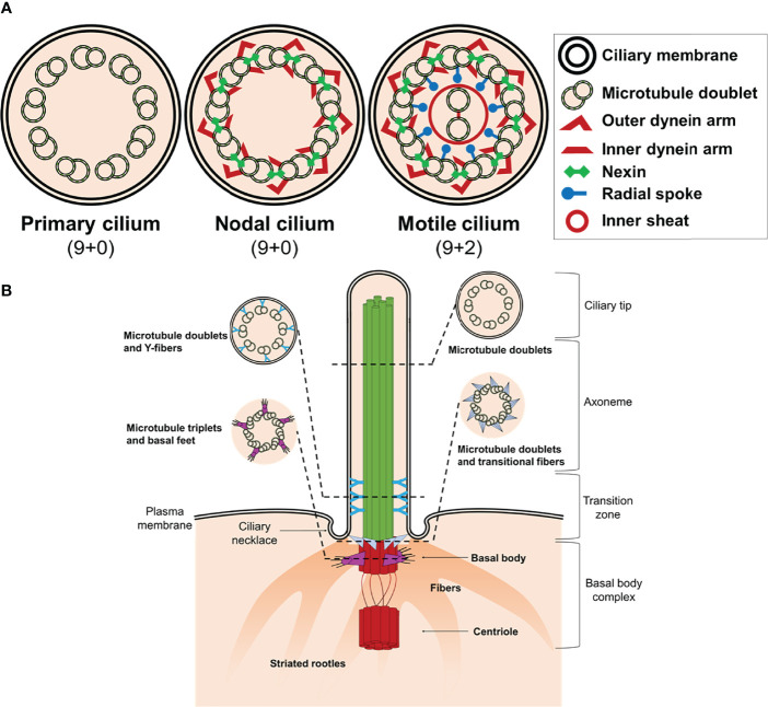
Figure 1.
Structure of cilium. (A) Schematic representation of an axoneme cross section from a primary cilium, nodal cilium, and motile cilium. The axoneme cilium is composed of nine outer doublets of microtubules surrounding a central pair (9 + 2). The axoneme is ensheathed by a ciliary membrane. Inner and outer dynein arms, nexin, and radial spokes are responsible to link microtubules and form a cylindrical structure. (B) Schematic diagram of a typical non-motile primary cilium. The primary cilium is divided into the ciliary tip, the membrane bound axoneme extending from surface, the transition zone, and the basal body complex. The ciliary tip ends contain signaling molecules and can undergo morphological changes in response to signaling processes. The axoneme is the structural core of a cilium. The transition zone converts the triplet microtubular structure of the basal body into the axonemal doublet structure. The ciliary pocket (necklace) is an invagination of the plasma membrane at the root of cilium. The basal body complex comprises the basal body and its centriole. In most quiescent cells, the centrioles move to the apical plasma membrane and the basal body (mother centriole) functions as the microtubule-organizing centre to nucleate the axonemal microtubules. The centriole (daughter centriole) remains perpendicular to the basal body.

