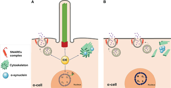
Figure 5.
Non-proteolytic functions of IDE in α-cells. (A) The abundance of IDE in pancreatic α-cells is relevant for maintaining several cellular functions, such as glucagon secretion, cytoskeletal organization, and ciliogenesis, while cells are maintained in a quiescent state. (B) Deletion of IDE in mouse α-cells revealed multiple phenotypes, such as hyperglucagonemia, hyperplasia, and hypertrophy, suggesting different non-proteolytic functions of IDE in these cells. Lack of primary cilia along with increased proliferation, in Ide depleted α-cells, provide direct functional evidence for the involvement of cilia in α-cell proliferation. Likewise, loss of IDE causes α-synuclein aggregation, which might underlie the absence of cilia, cytoskeletal alterations, and augmented SNARE proteins of the secretory machinery. This figure was created using Servier Medical Art (available at https://smart.servier.com/).

