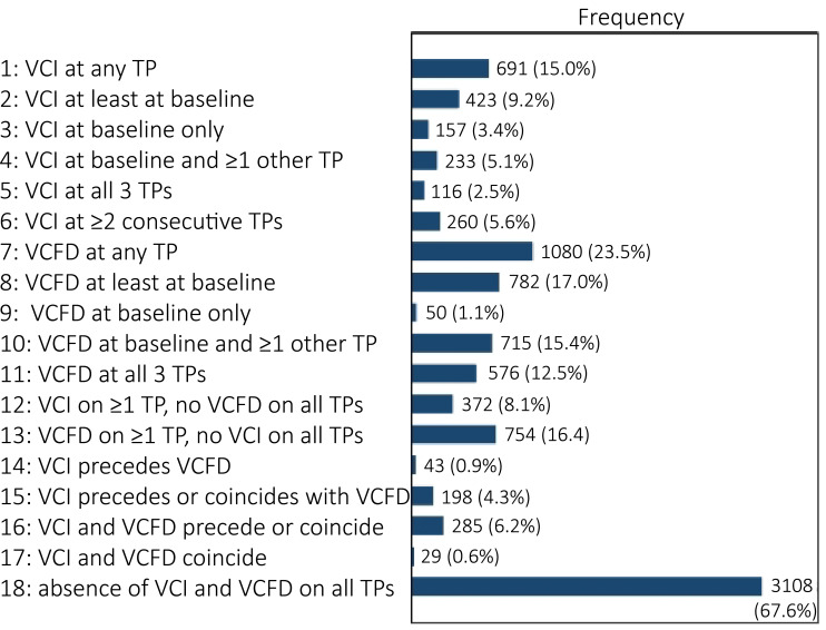
Figure 1.
Observed frequencies of patterns of MRI lesions. Patterns over time were studied in a total of 4600 vertebral corners. Patterns are deemed present if ≥2 out of 3 MRI readers scored the pattern over time. The patterns are not all mutually exclusive. For example, of the 4600 corners, 116 had presence of VCI at all timepoints (pattern 5). These corners are all included in the 691 corners with presence of VCI on at least one timepoint (pattern 1), and presence of VCFD is disregarded in these patterns. TP, timepoint; VCFD, vertebral corner fat deposition; VCI, vertebral corner inflammation.

