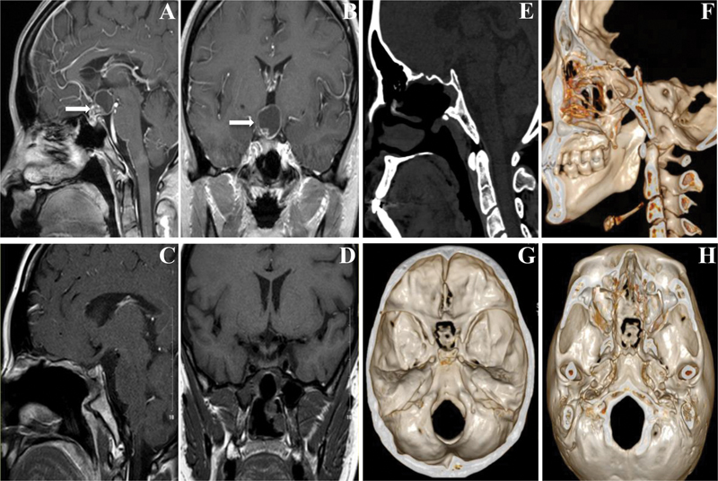
Fig. 2.

Representative images before and after surgery. ( A,B ) MRI results before surgery ( white arrows suggest the tumor ); ( C,D ) MRI results after surgery show complete resection of the tumor and good healing of the nasoseptal flap 1 month postoperatively; ( E–H ) 3D reconstruction of the skull base 1 year after surgery shows that bone window healed well using in situ bone flap and significant bone absorption was not seen. 3D, three-dimensional: MRI, magnetic resonance imaging.
