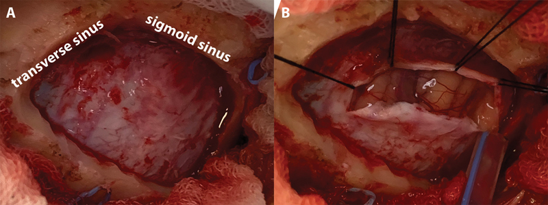
Fig. 2.

Craniotomy and cerebrospinal fluid drainage. ( A ) The junction of the transverse and sigmoid sinus is visualized. Craniotomy was not required to extend too inferiorly to approach the cisterna magna. ( B ) The cerebellum does not bulge through the craniotomy window even before cerebrospinal fluid release from the lateral medullary cistern.
