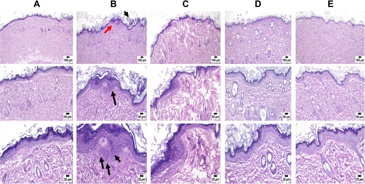
Figure 8.
Microscopic examination of skin samples from (A) control group revealing normal histology of skin, (B) positive control showing histopathological alterations mainly in the epidermis. Hyperkeratosis and acanthosis (black arrows) are manifested by thickening in the epidermal layer (red arrow). The epidermis contained numerous shrunken cells known as sunburn cells near the basal cell layer (3 black arrows), (C) Vit C-loaded NE showing apparently normal skin with mild inflammatory cells infiltration, (D) collagen-loaded NE showing apparently normal skin, and (E) a mixture of collagen and Vit C-loaded NEs revealing skin free from any detectable alterations.

