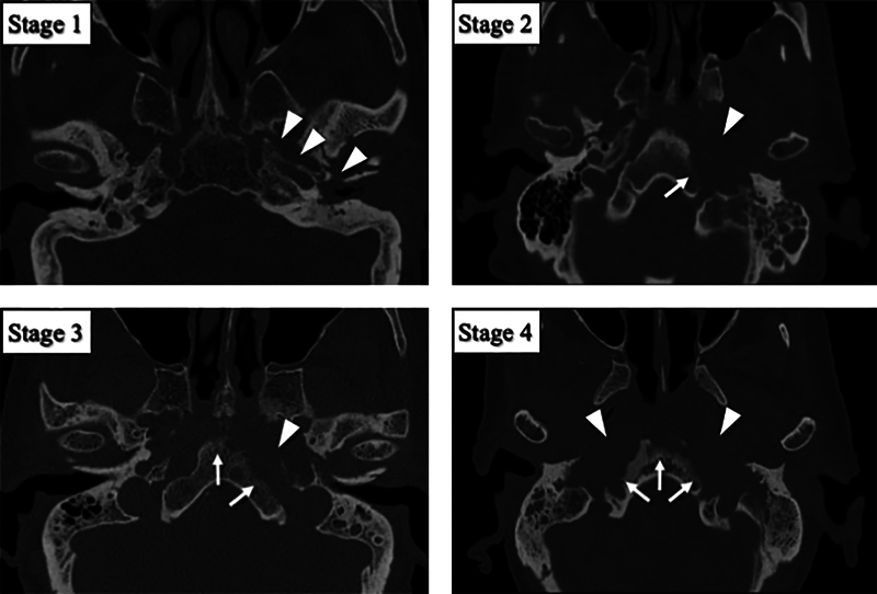
Fig. 1.

HRCT-based stages of otogenic skull base osteomyelitis. HRCT images were classified into four stages according to the cortical bone destruction. Arrow heads indicate areas of cortical bone destruction in the petrous portion of temporal bone. Arrows indicate areas of cortical bone destruction in the clivus. HRCT, high-resolution computed tomography.
