Fig. 3.

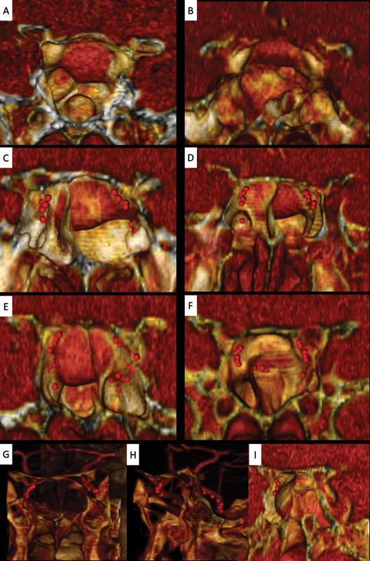
Examples of internal carotid artery (ICA) reconstruction based on OSIRIX. ( A ) and ( B ) With a good sphenoidal sinus pneumatization, the ICA prominences are naturally well demarcated without using region of interest (ROI) points. ( C–F ) Examples of cases where the ICAs are not clearly seen with the simple three-dimensional (3D) reconstructions because they are covered by any septum or are severely displaced by the tumor. The use of red ROI points previously marked in the two-dimensional axial CTA slices and automatically represented in our three-dimensional model allowing us to predict the position of the ICA preoperatively. Some anatomical variations can be identified beforehand, like a medialization of the ICA (i.e., right ICA of case F). ( G–I ) represents a macroadenoma with Knops 4 invasion of the left cavernous sinus. The ICA is marked by red ROI points and by using a combination of coronal and sagittal cuts and slight changes in the threshold or transparency of our 3D reconstruction, we are able to identify its complete course.
