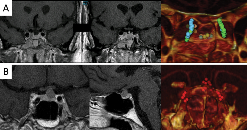
Fig. 5.

Examples of cases where the tumor is not in direct contact with the sellar floor. The use of region of interest points marking the tumor and changes in threshold/transparency allow us to predict the location of the tumor. ( A ) Case of a patient with acromegaly showing two separated tumoral nodules: one in the center of the pituitary gland and the other in contact with the right cavernous sinus behind the parasellar segment of the right internal carotid artery. ( B ) Case of a patient with Cushing disease showing a microadenoma close to the pituitary stalk. LHP, ---; RFA,---
