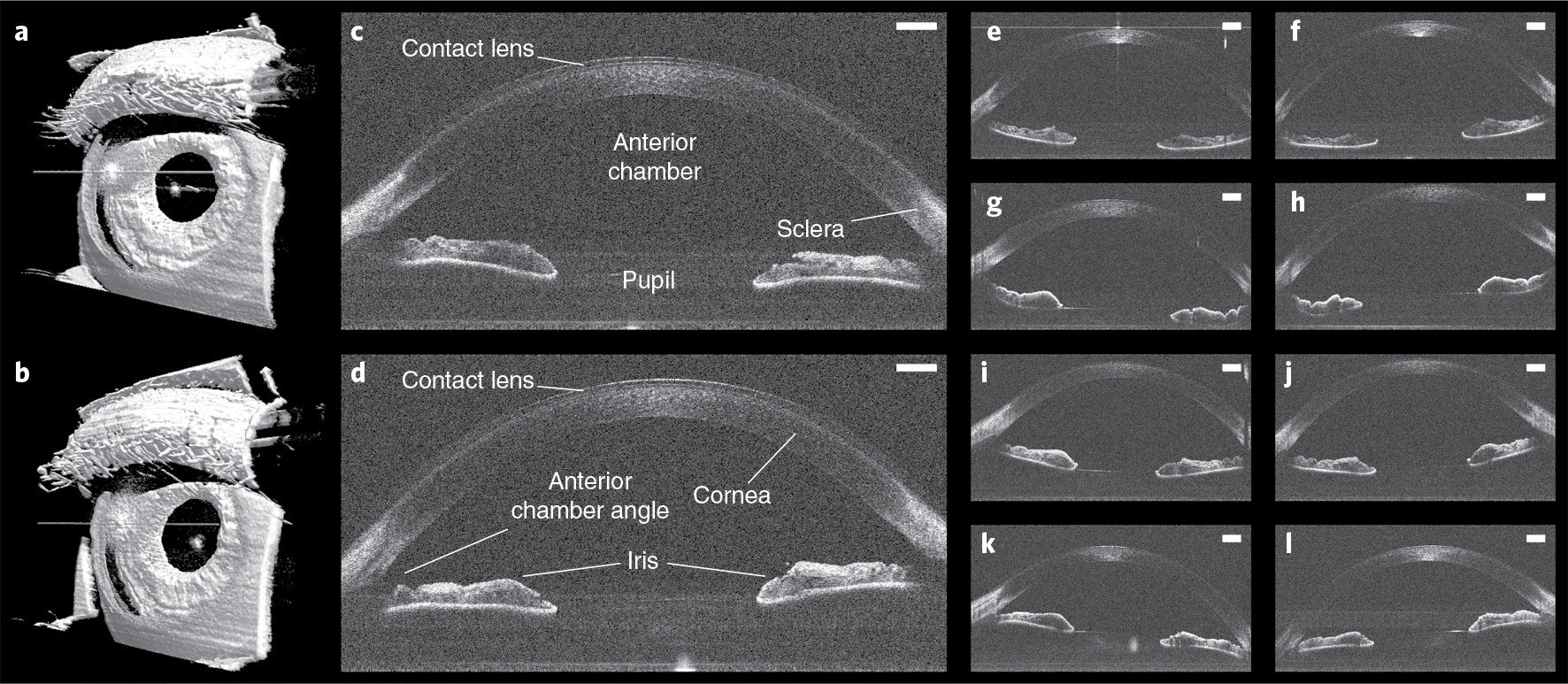
Fig. 3 |. Anterior imaging results in freestanding individuals.

a,b, Left (a) and right (b) 800 × 800 × 1,376 voxel anterior segment volumes obtained in a freestanding participant with automatic OCT imaging and registration in post-processing. c,d, Corresponding left (c) and right (d) anterior segment un-averaged B-scans from the same participant, revealing contact lens and relevant ocular anatomy. Scale bars, 1 mm. e–l, Left (e,g,i,k) and right (f,h,j,l) anterior segment un-averaged B-scans obtained with automatic OCT imaging from four additional participants, demonstrating reproducibility across participants. Scale bars, 1 mm.
