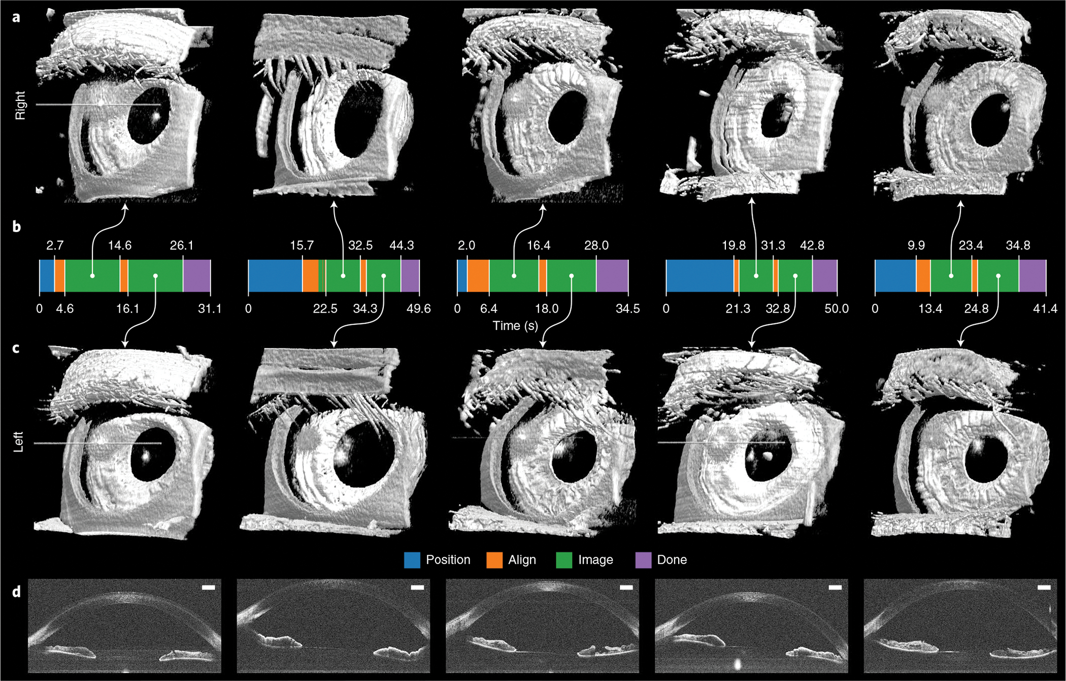
Fig. 5 |. Autonomous anterior segment results in freestanding individuals.

a,c, Paired right (a) and left (c) 800 × 200 × 1,376 voxel anterior segment volumes obtained with fully autonomous OCT imaging and registration in post-processing. Each right–left pair is from the same individual and imaging session. These scans captured the full cornea and iris surfaces, except those portions that the superior eyelid covered. Eyelashes are seen facing towards the eye in some volumes due to their large axial extent that produced wraparound complex-conjugate OCT artefacts. Raw volumes are shown in Supplementary Fig. 1. b, Autonomous system mode from initial to last participant detection. For all participants, the system reliably performed 10 s of imaging per eye after aligning with the right and then the left eye for a total session time of under 50 s. Moreover, the system recovered from eye-tracking loss, as seen with the second participant, and subsequently imaged for an uninterrupted 10 s. d, Un-averaged B-scans from volumes in c, revealing the cornea, anterior chamber, iris, angle of cornea and pupil as illustrated in Fig. 3. Scale bars, 1 mm.
