
Figure 4.
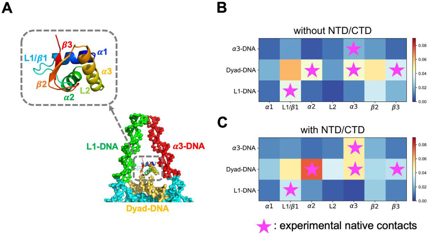
H1 disordered domains regulate and stabilize GH1-DNA binding interface. GH1 and nearby DNA structures are represented in (A), where colors represent different protein or DNA regions. (B-C) GH1-DNA contact maps without and with H1 NTD/CTD. Horizontal and vertical axes represent GH1 and DNA regions respectively. Color code from blue to red indicates the contact probability of a protein-DNA beads pair in this region from low to high. Purple stars represent the experimental “native contact regions”, where more than two residues in this GH1 region are close to a DNA region.