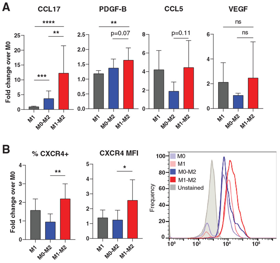
FIGURE 5. Differential protein expression in M1→M2 Mϕs.
(A) Protein secretion of selected highly-expressed M1→M2 genes. ELISA analysis of Mϕ-conditioned media. Data are represented as mean ± SD of fold change over M0 control. One-way ANOVA with Tukey’s post hoc conducted on log-transformed data, n = 4-11 donors, **P < 0.01, ***P < 0.001, ****P < 0.0001. (B) Flow cytometry analysis of CXCR4 surface expression. Data are represented as mean ± SD of fold change over M0 control. One-way ANOVA with Tukey’s post hoc conducted on log-transformed data, n = 7 donors, *P < 0.05, **P < 0.01. Representative histogram depicts one donor

