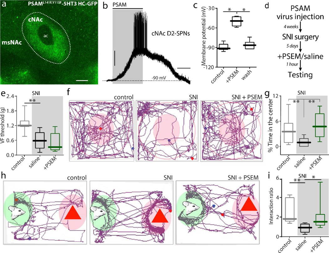
Figure 3. Chemogenetically activating cNAc d2SPNs relieved anxiety and social recognition deficits in SNI mice.

a, In AAV-PSAML141F,Y115F-5HT3 HC-GFP transduced A2A::Cre mice, GFP fluorescence was exclusively expressed in cNAc. b-c, PSEM89S induced firing in PSAM-5HT3-GFP expressing cNAc d2SPNs from A2a-Cre mice (n = 7 neurons from 5 mice per group in c, Wilcoxon test with W = 28, p = 0.0156 for PRE versus SPEM and W = 28, p = 0.0156 for PSEM versus WASH). d, The timeline for behavioral tests in the PSAM-5HT3 infected mice (n = 7 in Sham, n = 5–6 in Saline-SNI, n = 8 in PSEM-SNI; Mann–Whitney U test for data statistics). e, Activating cNAc d2SPNs by i.p. injection of PSEM89S had no effect on SNI-induced tactile allodynia (U = 1, p = 0.0051 for Sham versus Saline-SNI; U = 15.50, p = 0.5462 for Saline-SNI versus PSEM-SNI).f-g, In open field test, PSAM-5HT3 expressing SNI mice receiving PSEM89S treatment spent more time in the center zone of open field than those receiving saline treatment (U = 2, p = 0.0062 for Sham versus Saline-SNI; U = 2, p = 0.0062 for Saline-SNI versus PSEM-SNI). h-i, In social recognition task, PSEM89S treatment abolished the deficits in social recognition associated with SNI (n = 7 in Sham, n = 6 in Saline-SNI, n = 8 in PSEM-SNI; U = 1, p = 0.0023 for Sham versus Saline-SNI; U = 6, p = 0.02 for Saline-SNI versus PSEM-SNI). Data are presented as whisker box plots displaying median, lower and upper quartiles, and whiskers representing minimum and maximum of the data.
