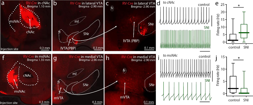
Figure 7. SNI differentially modulated VTA dopaminergic innervation of cNAc and msNAc.
a-c, After the injection of RV-Cre into the cNAc of an Ai14-tdTomato-flox mouse, tdTomato signal was restricted to the cNAc (a) and retrogradely labeling was detected in the lateral VTA (lVTA, b-c). Robust tdTomato expression was routinely detected in the VTA, but no attempt was made to rigorously estimate the percentage of the neurons labeled. d-e, The intrinsic neuronal excitability of VTA-DA neurons innervating the cNAc was upregulated following SNI (n = 17 neurons from 6 mice per group, U = 76, p = 0.0173). d, Sample traces of VTA neurons innervating cNAc recorded from Sham and SNI slices. f-h, The VTA neurons projecting to NAc medial shell were dominantly located in medial VTA (mVTA) of the rostral midbrain. i-j, Medial VTA neurons projecting to msNAc from SNI mice had slower firing than that from Sham mice (n = 15 neurons from 6 mice per group, U = 61, p = 0.0319). ml: medial lemniscus; PBP: parabrachial pigmented nucleus of the VTA; SNr: substantia nigra, reticular part; VS: ventral subiculum; VTA: ventral tegmental area. Data are analyzed by Mann–Whitney U test and presented as whisker box plots displaying median, lower and upper quartiles, and whiskers representing minimum and maximum of the data.

