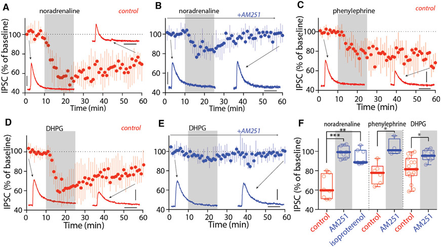
Figure 6. Activation of α1ARs and mGluR-I induced LTD at CeA synapses on lateral PBN.
(A and B) NA (10 μM, A) induced LTD of CeA IPSCs, which was blocked by AM251 (B).
(C) Activation of α1ARs with phenylephrine (Phe, 10 μM) induced LTD of CeA IPSCs.
(D and E) Activation of mGluR-I with DHPG (100 μM, D) induced LTD of CeA IPSCs, which was blocked by AM251 (E).
(F) Summary showing activation of α1ARs with Phe and mGluR-I with DHPG, but not βARs with isoproterenol, induced eCB-LTD at CeA synapses on PBN.
Scale bars: 40 ms and 100 pA in (A)–(E). Statistics: *p ≤ 0.05, **p ≤ 0.005, ***p ≤ 0.001, Kruskal Wallis with Dunn’s correction (left), Mann-Whitney U test (middle and right). Also see Table S1.

