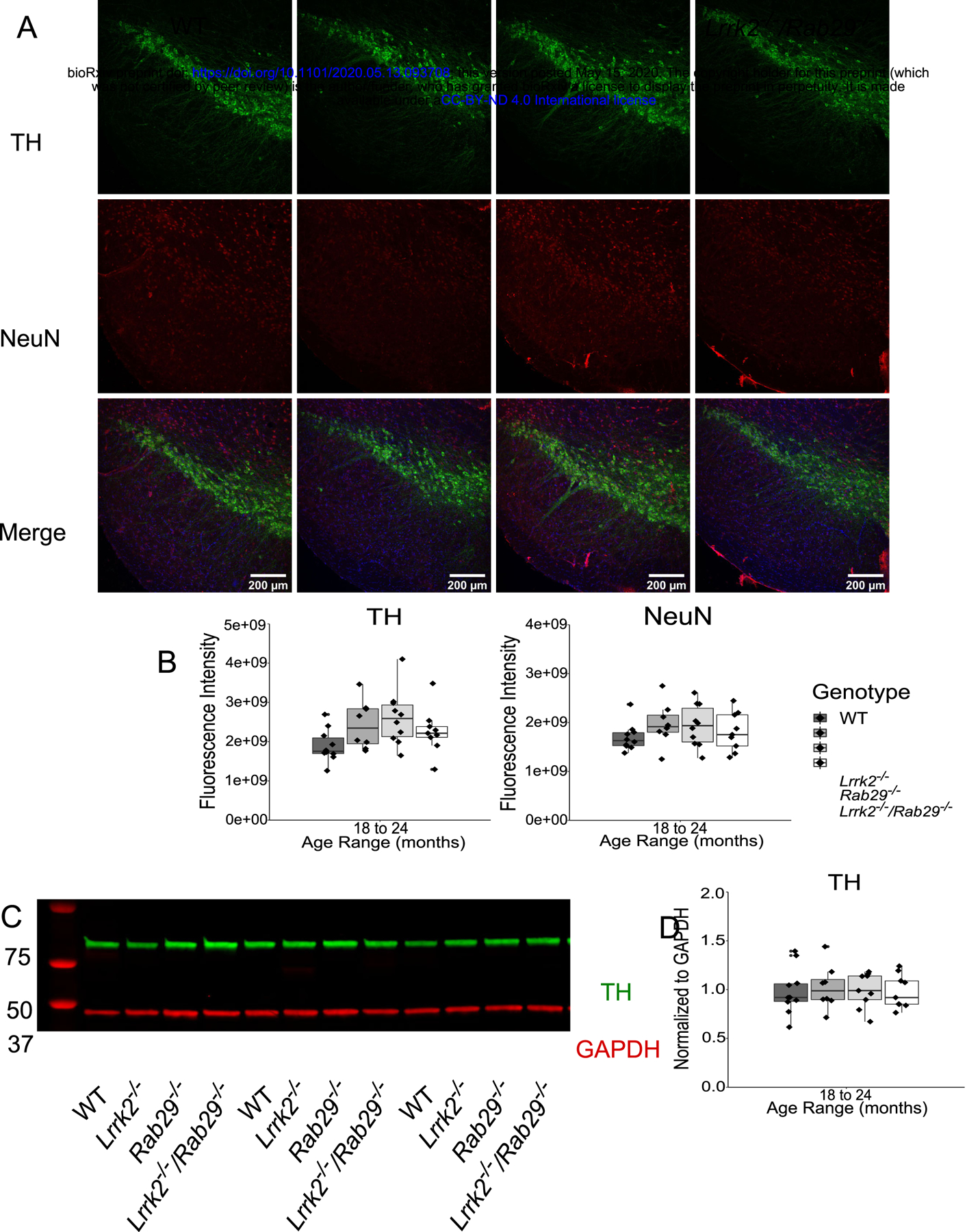
Figure 4.

Postmortem analyses of dopamine loss in the SNc and striatum of 18 – 24 month single and double knockout mice. Immunohistochemistry images of SNc stained for TH (green), NeuN (red), and Hoechst (blue; A). Genotype differences in TH (B) and NeuN (C) mean fluorescence intensity were quantified. Western blot analysis of striatal tissue expression of TH and GAPDH (D). Striatal TH was normalized to GAPDH and quantified for genotype differences (E).
