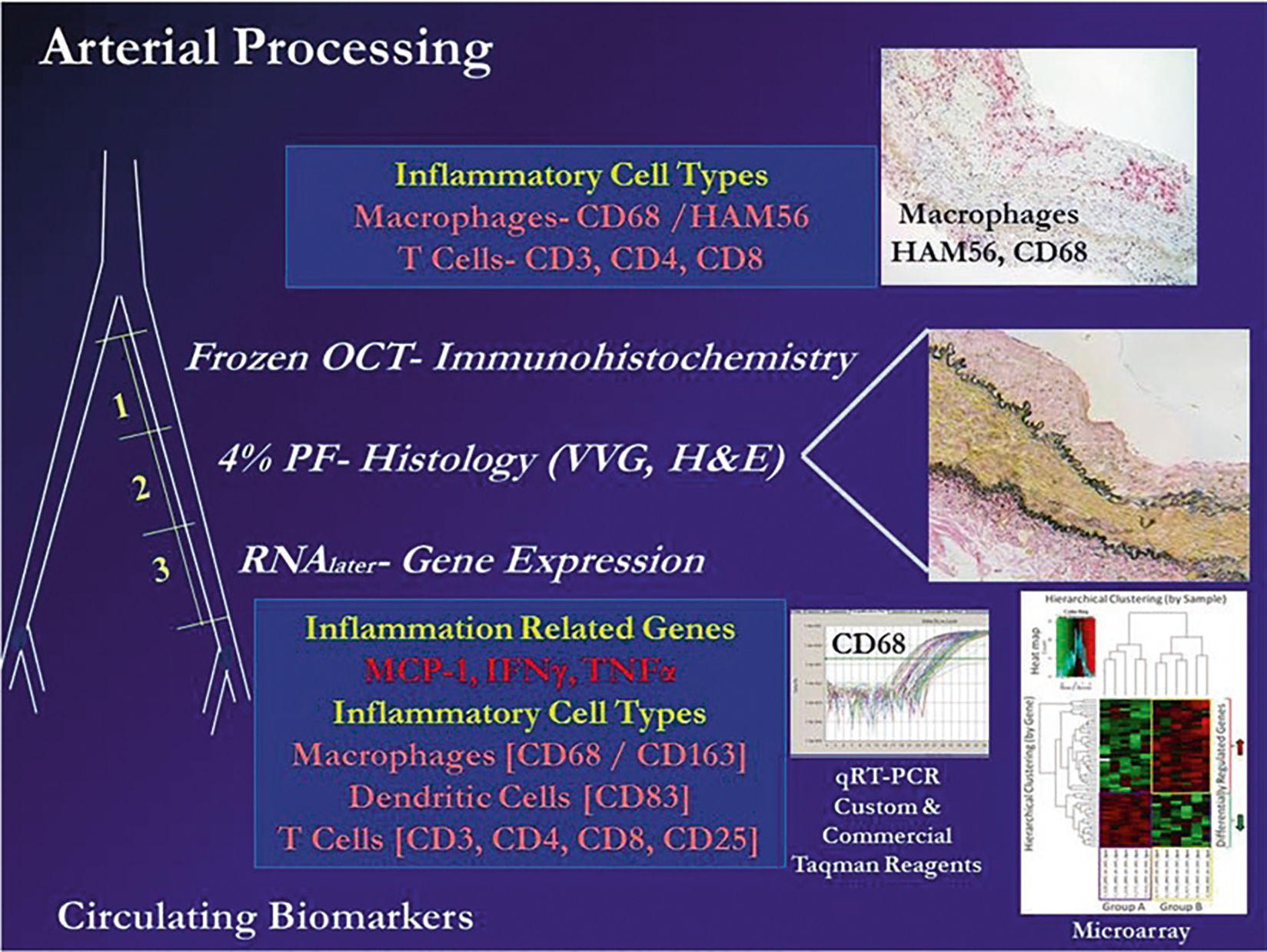
Fig. 2.

Diagram of approaches to evaluation of cellular, morphologic, and molecular phenotypes in an iliac artery biopsy or another arterial specimen obtained ante or post mortem in response to diet and interventions. A biopsy of the iliac artery can be obtained surgically and prepared for immunohistochemistry (image 1, cryopreserved in OCT embedding medium), histology (image 2, fixed with 4 % paraformaldehyde, moved to 70 % alcohol, and then embedded in paraffin), and for assessment of gene expression by qRT-PCR, microarray, or RNAseq (images 3 and 4, prepared in RNAlater® or slam frozen). Abbreviations: OCT Optimal Cutting Temperature freezing embedding medium, VVG Verhoeff-van Gieson stain, H and E hematoxylin and eosin. Macrophage markers CD68 and/or HAM56 (monoclonal antibody to human alveolar macrophages), CD163 (M2 marker hemoglobin-haptoglobin receptor), T cell markers CD3 (T cell lineage), CD4 (T helper cells), CD8 (NK cells), CD25 (Tregs). Dendritic cells CD83 [24, 31, 32]. In general, estrogen treatment initiated soon after the loss of estrogen (through ovariectomy) causes a 50–70 % inhibition of diet induced atherosclerotic plaque size in the coronary arteries on monkeys relative to untreated controls. Molecular and cellular responses to estrogen (reduced levels of arterial expression of inflammation and inflammatory cell associated markers) can be detected well before changes in lesion size can be observed; therefore, molecular approaches can significantly reduce sample sizes required for appropriate statistical power to see an effect
