
Figure 5:
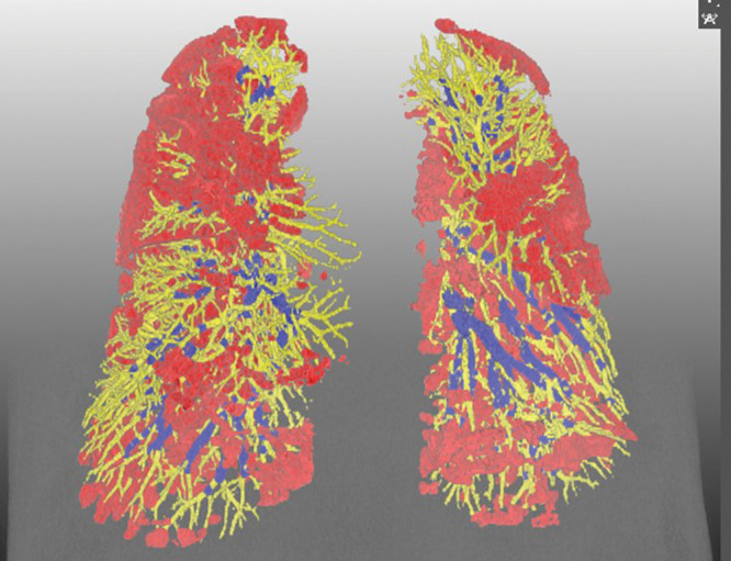
Representative three-dimensional chest CT image in a 52-year-old man with the Omicron variant of COVID-19 shows pneumonia evenly affecting lungs (pneumonia volume, 17.5%) and a preserved percentage of blood volume in intrapulmonary vessels, with a cross-sectional area less than 5 mm2 relative to the total pulmonary blood volume (51.5%). Blue vessels have a cross-sectional area of 5 mm2 or greater, and yellow vessels have a cross-sectional area less than 5 mm2. Red indicates COVID-19 pneumonia.