Figure 3.

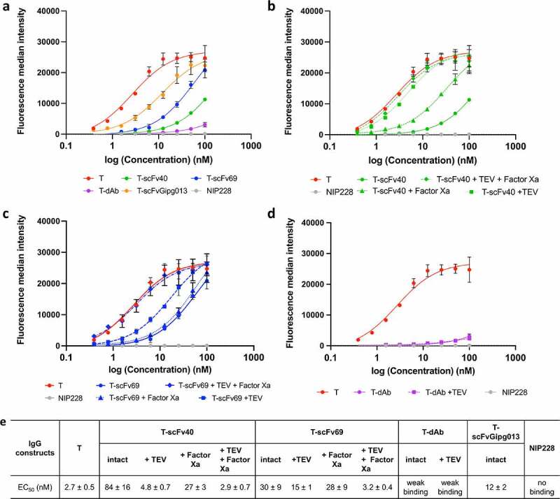
Fluorescence median intensity measured by flow cytometry with channel detecting the AlexaFluor 647 fluorophore signal from a secondary antibody. All experiments were performed in triplicate, the mean and the standard deviation are plotted. The data in panel a show the masking efficiency of the masked antibodies and control, whilst the data in panels b-d show the degree to which the masked antibodies can be activated by cleavage with proteases. a) Fluorescence median intensity of the secondary antibody showing the binding of the masked intact antibodies against HER2. b) Fluorescence median intensity of the secondary antibody showing the binding of the T-scFv40 constructs cleaved with TEV and/or Factor Xa to HER2, illustrating the degree of activation of the T-scFv40 upon cleavage. c) Fluorescence median intensity of the secondary antibody showing the binding of T-scFv69 to HER2 after cleavage with TEV and/or Factor Xa proteases. d) Fluorescence median intensity of the secondary antibody showing the binding of T-dAb to HER2 after cleavage of the linker with TEV protease. e) EC50 parameters obtained from the flow cytometry measurements, fitted to Equation 1.
Figure 3 Alt text: Figure made of five panels. In panels a to d, four graphs representing the binding (from fluorescence intensity) of intact and cleaved masked to HER2 on SK-BR-3 cells is shown as a function of antibody concentration. In panel a, the binding curves of intact antibodies to HER2 are shown: naked trastuzumab binds the most, followed by T-scFvGipg013, T-scFv69, T-scFv40, T-dAb and NIP228. In panel b, the binding of intact and cleaved T-scFv40 to HER2 is shown: naked trastuzumab, T-scFv40 + TEV and T-scFv40 + TEV+ Factor Xa bind the most and equally, followed by T-scFv40 + Factor Xa, followed by T-scFv40 intact. In panel c, the binding of intact and cleaved T-scFv69 to HER2 is shown: naked trastuzumab, T-scFv69 + TEV + Factor Xa bind the most and equally, followed by T-scFv69 + TEV, and followed by T-scFv69 intact and T-scFv69 + Factor Xa which bind equally. In panel d, the binding of intact and cleaved T-dAb to HER2 is shown: naked trastuzumab binds the most, T-dAb intact and cleaved bind equally and very little. The panel e is a table with the EC50 fittings of the curves.
